
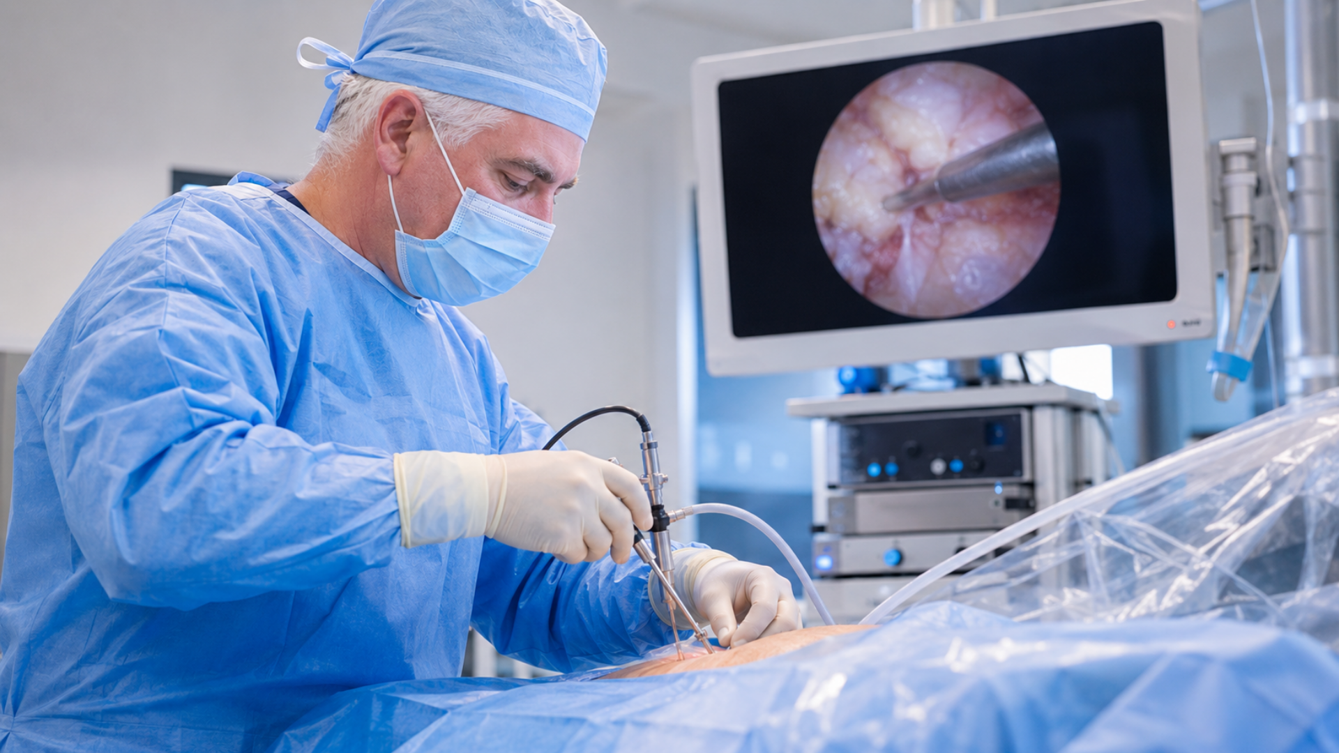
What Is Endoscopic Spine Surgery in Lewisville TX?
Endoscopic spine surgery is an advanced, minimally invasive procedure that treats nerve compression in the spine through an incision smaller than a quarter-inch. A neurosurgeon inserts a thin tube equipped with a high-definition camera and specialized instruments directly to the source of the problem, then removes the tissue pressing on the nerve while watching a magnified live video feed. Scott C. Kutz, MD, a board-certified neurosurgeon at Minimally Invasive Neurosurgery of Texas, performs endoscopic spine surgery for appropriately selected patients at the practice’s Lewisville and Plano, Texas locations, serving patients seeking endoscopic spine surgery Lewisville TX, and minimally invasive endoscopic spine surgery Lewisville TX.
Unlike traditional open spine surgery, which requires a large incision and separating back muscles from the spine, endoscopic surgery works through the natural spaces between muscles. The endoscope, roughly the width of a pencil, provides a clear, magnified view of the spinal canal without requiring the surgeon to cut through muscle or remove stabilizing bone structures. Because the lamina, facet joints, and ligaments remain intact, the procedure is classified as motion-preserving. Patients keep their natural range of spinal movement after surgery.
Most endoscopic spine procedures are performed on an outpatient basis, meaning patients go home the same day. Many cases can be completed under local anesthesia with sedation rather than general anesthesia, which reduces anesthesia-related risk for older patients or those with other medical conditions. Dr. Kutz completed advanced Endoscopic Spine Surgery Level 1-2 Workshop certification and applies these techniques as part of a broader commitment to minimally invasive spine surgery at Minimally Invasive Neurosurgery of Texas.
What Conditions Can Endoscopic Spine Surgery Treat?
Endoscopic spine surgery treats conditions in which a specific spinal structure compresses or irritates a nerve. The procedure works best when imaging clearly identifies the source of the problem and the patient’s symptoms match that finding. Conditions commonly treated with this technique include:
Herniated discs are the most common reason patients undergo endoscopic surgery. When the soft inner material of a spinal disc pushes through a tear in the outer wall, it can press directly on a nearby nerve root. The surgeon uses the endoscope to locate and remove only the disc fragment causing the compression, leaving the healthy portion of the disc intact.
Lumbar herniated discs causing sciatica respond particularly well to endoscopic discectomy. The procedure relieves radiating leg pain, numbness, and tingling caused by nerve root compression in the lower back.
Endoscopic surgery can also address cervical herniated discs causing arm pain, numbness, or weakness in select cases, depending on the location and size of the herniation.
Foraminal stenosis occurs when the bony exit window where a nerve root leaves the spinal canal narrows and squeezes the nerve. Endoscopic foraminotomy widens this opening to relieve pressure without removing the spine’s stabilizing structures.
In select cases, surgeons can treat spinal stenosis endoscopically. When the narrowing is focal and the anatomy allows safe endoscopic access, the surgeon can decompress the affected nerve through the endoscope. More extensive or multilevel stenosis may require a different surgical approach.
Bone spurs (osteophytes) that develop from degenerative changes and press on nerves can be carefully removed through the endoscopic working channel.
Facet joint cysts that compress nerve roots can be drained or removed endoscopically.
Recurrent disc herniation after a prior surgery is treatable endoscopically in many cases. Because the endoscope works through a new, clean pathway, it avoids the scar tissue from the original operation.
Sciatica and radiculopathy caused by any of the above conditions often improve significantly after removing the source of nerve compression.
Not every spinal condition is appropriate for endoscopic treatment. Dr. Kutz evaluates each patient individually to determine whether the anatomy and diagnosis support an endoscopic approach or whether a different technique would produce a better outcome.
Who May Be a Candidate for Endoscopic Spine Surgery?
Candidacy for endoscopic spine surgery depends on several factors, including the specific diagnosis, the location of the problem, and how the patient’s symptoms match the imaging findings.
Patients who may benefit from endoscopic spine surgery typically share these characteristics:
Persistent radicular pain. Arm or leg pain that follows a nerve pathway, often described as sharp, burning, or electric, is the hallmark symptom. This pain results from a compressed or irritated nerve root and is what endoscopic decompression is designed to relieve.
Failed conservative treatment. Most spine specialists recommend trying nonsurgical options first. Physical therapy, anti-inflammatory medications, and epidural steroid injections often provide relief. Surgery becomes a reasonable consideration when these treatments have not improved symptoms after six to twelve weeks of consistent effort.
Imaging that confirms a structural cause. An MRI or CT scan must show a clear structural problem, such as a herniated disc or foraminal narrowing, that matches the patient’s symptoms. Dr. Kutz requires this clinical-radiologic correlation before recommending surgery. Imaging findings alone, without matching symptoms, do not make someone a surgical candidate.
Patients with higher surgical risk. Because endoscopic surgery can often be performed under local anesthesia with sedation, it may be a safer option for older adults or patients with medical conditions that increase the risk of general anesthesia.
Candidacy always requires a thorough in-person evaluation. Dr. Kutz reviews each patient’s imaging, medical history, and physical examination findings before determining whether endoscopic spine surgery is the right approach. To schedule an evaluation, contact Minimally Invasive Neurosurgery of Texas at (972) 244-3491.
Who May Not Be a Candidate?
Endoscopic spine surgery is not the right approach for every patient or every spinal condition. Understanding these limitations is an important part of making an informed decision.
Severe spinal instability. When the spine cannot maintain its normal alignment under load, the patient may need a stabilization procedure such as spinal fusion. Endoscopic surgery decompresses nerves but does not add structural support.
Advanced multilevel degenerative disease. Patients with significant degeneration affecting multiple spinal levels may need a more extensive surgical approach that addresses all affected areas in a single procedure.
Significant scoliosis or spinal deformity. Abnormal curvature can alter the anatomy in ways that limit safe endoscopic access to the affected nerve.
Anatomical variations. Some patients have bone structure or soft tissue configurations that make it difficult to create a safe working channel for the endoscope.
Conditions requiring instrumentation. If the treatment plan involves placing screws, rods, or an artificial disc, a different surgical approach is necessary.
Symptoms that do not match imaging. If a patient’s pain pattern does not match the structural findings on MRI or CT, surgery is unlikely to help. Dr. Kutz emphasizes the importance of accurate diagnosis before proceeding with any intervention.
Conservative treatment may still be appropriate. Some patients who have not yet completed a full course of physical therapy, medication management, or injection therapy may benefit from continuing those efforts before considering surgery.
How Is Endoscopic Spine Surgery Performed?
Understanding what happens during the procedure helps patients feel more confident and prepared. Here is a step-by-step overview of how endoscopic spine surgery works at Minimally Invasive Neurosurgery of Texas.
Preparation and anesthesia. The patient is positioned on the operating table, typically lying face down or on their side depending on the approach. Many endoscopic procedures use local anesthesia with IV sedation, which numbs the surgical area while keeping the patient comfortable but not fully unconscious. General anesthesia is used when the procedure or patient factors require it.
Creating the access point. The surgeon makes a single incision approximately 7 millimeters long, smaller than a thumbnail. Using real-time fluoroscopic (X-ray) guidance, the surgeon inserts a series of progressively larger dilators to gently separate the muscle fibers and create a working channel to the spine. This process pushes muscles aside rather than cutting through them.
Inserting the endoscope. A thin tubular cannula is placed through the working channel, and the endoscope is inserted through it. The endoscope contains a high-definition camera, a light source, and a continuous irrigation system that keeps the surgical field clear. The camera transmits a magnified view of the spinal structures to a monitor, giving the surgeon a direct and detailed view of the nerve, disc, and surrounding anatomy.
Performing the decompression. Using specialized micro-instruments passed through the endoscope’s working channel, the surgeon carefully removes the tissue causing nerve compression. This may involve removing a herniated disc fragment, trimming a bone spur, widening a narrowed foramen, or draining a cyst. The surgeon can directly visualize the nerve throughout the procedure, confirming full decompression before finishing.
Closure. The instruments and cannula are removed. The small incision typically requires a single stitch or adhesive strip. No drain is needed.
Immediate recovery. The patient moves to a recovery area for monitoring, usually for one to two hours. Most patients stand and walk before leaving the facility. Discharge typically occurs the same day.
Potential Benefits of Endoscopic Spine Surgery
Endoscopic spine surgery offers several advantages over traditional open procedures, though individual results depend on the specific condition treated and the patient’s overall health.
Smaller incision. The quarter-inch incision used in endoscopic surgery compares with the three- to six-inch incisions common in traditional open procedures. A smaller opening means less visible scarring and a lower risk of wound complications.
Reduced muscle and tissue disruption. Because the surgeon works through the natural spaces between muscles rather than cutting or detaching them, the back’s stabilizing structures remain intact. This is a significant difference from open surgery, where muscles must be stripped away from the spine to gain access.
Motion preservation. The lamina, facet joints, and spinal ligaments stay in place during endoscopic decompression. This preserves the spine’s natural flexibility and reduces the risk of adjacent segment disease. In this condition, the spinal levels near a rigid surgical site break down faster due to increased stress.
Outpatient procedure. Most patients go home the same day. This avoids the risks, costs, and inconvenience of an overnight hospital stay.
Faster recovery. Patients often return to desk work within one to two weeks and resume physically demanding activities within six to twelve weeks, compared to significantly longer timelines with traditional surgery.
Reduced post-operative pain. Less tissue disruption translates to less post-surgical pain and a lower need for narcotic pain medication.
Lower anesthesia risk. The option to use local anesthesia with sedation rather than general anesthesia reduces risk for patients with heart, lung, or other conditions that make general anesthesia more dangerous.
Lower infection risk. Smaller incisions have a lower rate of surgical site infection compared to larger open wounds.
These benefits represent what clinical evidence and patient outcomes generally demonstrate. Individual results vary, and no surgical procedure guarantees a specific outcome. Dr. Kutz discusses expected benefits and realistic expectations with each patient before surgery.
Risks and Considerations
Every surgical procedure carries risk. Patients considering endoscopic spine surgery should understand both general and procedure-specific risks.
General surgical risks include infection, bleeding, and adverse reactions to anesthesia or medications. These risks exist with any operation but are generally lower with minimally invasive approaches due to the smaller incision and reduced tissue disruption.
Nerve injury is a rare but serious potential complication. The surgeon works in proximity to spinal nerves throughout the procedure. Advanced visualization through the endoscope helps minimize this risk, but it cannot be eliminated.
Incomplete decompression may occur if anatomy limits the endoscope’s ability to reach all tissue compressing the nerve. In some cases, a second procedure or a conversion to a different surgical technique may be necessary.
Recurrent disc herniation can happen after any discectomy, including endoscopic. Disc material can re-herniate at the same level, or a new herniation can develop at a different level over time. Published data suggest recurrence rates are comparable between endoscopic and traditional microdiscectomy.
Dural tear (CSF leak) is an uncommon complication where the thin membrane covering the spinal cord and nerves is inadvertently punctured. This can cause a headache and typically heals on its own or with a minor follow-up procedure.
Published complication data. A 2025 retrospective study of 1,000 patients who underwent transforaminal endoscopic spine surgery reported complication rates of approximately 1.5 to 3.4 percent, comparable to or lower than traditional open microsurgery (Tornatore et al., Diagnostics, 2025). Pain scores in that study decreased from a mean of 8.2 before surgery to 2.1 at twelve months after surgery.
Dr. Kutz reviews all potential risks with each patient during the consultation and answers questions about how these risks apply to the individual’s specific situation.
Recovery After Endoscopic Spine Surgery
Recovery after endoscopic spine surgery is generally faster than recovery from traditional open spine surgery. However, the exact timeline depends on the procedure performed, the condition treated, the patient’s age, and overall health.
Day of surgery. Most patients stand and walk within one to two hours after the procedure. The clinical team monitors vital signs and confirms stable neurological function before discharge. Same-day discharge is typical.
First 24 to 48 hours. Rest at home with short, frequent walks. Ice therapy applied to the incision area helps manage swelling. Most patients manage discomfort with over-the-counter pain medication or a short course of prescribed medication. Heavy narcotics are usually unnecessary.
Weeks one to two. Patients gradually increase their walking distance. Light household activities are generally safe. Many patients with desk jobs return to work during this period, as long as they can stand and stretch periodically. Driving is usually safe once the patient is no longer taking any pain medication that impairs alertness.
Weeks three to six. Activity levels increase further. Patients often resume most daily routines. Low-impact exercise such as walking and swimming may begin with clearance from Dr. Kutz.
Six to twelve weeks. Gradual return to more demanding physical activities. Patients with physically demanding jobs typically return to full duty during this window. Contact sports and heavy lifting require specific clearance.
Follow-up care. Dr. Kutz schedules post-operative visits to monitor healing, review imaging when indicated, and clear patients for progressive activity milestones. The follow-up schedule is tailored to each patient’s recovery.
When to seek immediate medical attention. Contact the office or go to the emergency room if you experience sudden or progressive weakness in your arms or legs, loss of bladder or bowel control, high fever with severe back pain, or worsening pain that is not controlled by your prescribed medication. These symptoms are rare but require prompt evaluation.
Recovery timelines above are general estimates. Every patient heals differently, and Dr. Kutz provides individualized guidance based on the specific procedure and the patient’s progress.
Endoscopic Spine Surgery vs. Traditional Open Spine Surgery
Patients often want to understand how endoscopic spine surgery compares to traditional open approaches. Both methods can effectively decompress nerves and relieve pain. The difference lies in how the surgeon accesses the spine and how that access affects surrounding tissue.
Incision and muscle disruption. Traditional open surgery typically requires a three- to six-inch incision. The surgeon must separate or detach the paraspinal muscles from the spine to see the surgical area. This muscle disruption is a major contributor to post-operative pain and prolonged recovery. Endoscopic surgery uses an incision under a quarter-inch and works between muscle fibers, avoiding significant muscle damage.
Hospital stay. Open decompression procedures often require one to three days in the hospital. Endoscopic procedures are typically same-day outpatient surgeries.
Recovery time. Open surgery patients often need four to eight weeks before returning to desk work and three to six months before resuming full physical activity. Endoscopic patients commonly return to desk work in one to two weeks and full activity in six to twelve weeks.
Anesthesia. Open surgery almost always requires general anesthesia. Many endoscopic procedures can be performed under local anesthesia with sedation, which carries lower cardiovascular and pulmonary risk.
Spinal stability. Open laminectomy removes bone (the lamina) to access the spinal canal, which can reduce spinal stability and sometimes leads to the need for fusion. Endoscopic decompression preserves the lamina and facet joints, maintaining the spine’s natural structure.
Clinical outcomes. A comprehensive review published in The Spine Journal in March 2026 (Kwon and Moon) confirmed that endoscopic lumbar discectomy produces pain relief and functional improvement equivalent to traditional microdiscectomy, with the added benefits of smaller incisions, less soft tissue disruption, reduced blood loss, and shorter hospital stays.
When traditional surgery may be the better choice, not every patient or condition is suited for endoscopic treatment. Severe spinal instability, multilevel stenosis requiring broad decompression, complex spinal deformity, and conditions requiring fusion or instrumentation are generally better addressed through traditional or other minimally invasive approaches. Dr. Kutz evaluates each case individually and recommends the approach most likely to produce the best long-term outcome for that patient.
Who Performs Endoscopic Spine Surgery in North Texas?
Scott C. Kutz, MD, is a board-certified neurosurgeon and Fellow of the American Association of Neurological Surgeons who performs endoscopic spine surgery at Minimally Invasive Neurosurgery of Texas. Dr. Kutz earned his medical degree from the University of Illinois College of Medicine and completed his neurosurgical residency at Louisiana State University in New Orleans, followed by a fellowship in stereotactic and functional neurosurgery at Rush University Medical Center in Chicago. He has performed over 6,000 successful spine surgeries and consulted with more than 25,000 patients over his 27-year career.
Dr. Kutz has published peer-reviewed content on Spine-Health, including articles on sacroiliac joint fusion and how posture affects lumbar herniated discs. He holds Endoscopic Spine Surgery Level 1-2 Workshop certification and integrates endoscopic techniques alongside the Globus Excelsius GPS Robotic Navigation Platform and Augmedics augmented reality system used at the practice.
Minimally Invasive Neurosurgery of Texas evaluates patients at two locations in North Texas:
Lewisville office: 1850 Lakepointe Drive, Suite 500, Lewisville, TX 75057 Plano office: 5465 Legacy Drive, Suite 650, Plano, TX 75024
The practice also serves patients from surrounding communities, including Frisco, Flower Mound, Denton, McKinney, and Allen.
Patients seeking endoscopic spine surgery Lewisville TX, can call (972) 244-3491 to schedule a consultation and learn whether this approach is appropriate for their condition, or visit the contact page.
Frequently Asked Questions
What is endoscopic spine surgery? Endoscopic spine surgery is a minimally invasive procedure that uses a small camera and specialized instruments inserted through an incision smaller than a quarter inch. The surgeon watches a magnified live video feed to locate and remove tissue compressing a spinal nerve. Most procedures are outpatient, and patients go home the same day.
Is endoscopic spine surgery the same as minimally invasive spine surgery? Endoscopic spine surgery is one type of minimally invasive spine surgery, but the two terms are not interchangeable. Minimally invasive spine surgery is a broad category that includes many techniques using smaller incisions, such as tubular retractor-based procedures and robotic-assisted surgery. Endoscopic surgery specifically uses an endoscope, a thin tube with a camera, to operate through the smallest possible opening.
What conditions can endoscopic spine surgery treat? Common conditions treated endoscopically include herniated discs, foraminal stenosis, select cases of spinal stenosis, bone spurs compressing nerves, facet joint cysts, and recurrent disc herniations. The procedure works best when imaging confirms a specific structural problem that matches the patient’s symptoms.
Am I a candidate for endoscopic spine surgery? Candidates typically have persistent arm or leg pain caused by a compressed nerve, have tried conservative treatments such as physical therapy and injections without adequate relief, and have imaging that confirms a structural cause matching their symptoms. Candidacy requires an individualized evaluation by a spine surgeon. Not all conditions or anatomies are suited for endoscopic treatment.
Is endoscopic spine surgery performed as an outpatient procedure? Yes, in most cases. Patients typically go home within a few hours of the procedure. Same-day discharge is standard for most endoscopic spine surgeries performed at Minimally Invasive Neurosurgery of Texas.
How long does endoscopic spine surgery take? Most endoscopic procedures take 45 to 90 minutes, depending on case complexity and the number of levels treated. The exact duration varies by patient.
What is the recovery time after endoscopic spine surgery? Many patients return to desk work within one to two weeks and resume full physical activity within six to twelve weeks. Recovery varies based on the specific procedure, the condition treated, patient age, and overall health. Dr. Kutz provides a personalized recovery plan for each patient.
What are the risks of endoscopic spine surgery? Risks include infection, nerve injury, incomplete decompression, recurrent disc herniation, and dural tear. Published complication rates for endoscopic spine surgery are approximately 1.5 to 3.4 percent in large patient cohorts, comparable to or lower than traditional open microsurgery. Dr. Kutz discusses all risks during the pre-operative consultation.
Who performs endoscopic spine surgery in Lewisville and Plano TX? Scott C. Kutz, MD, a board-certified neurosurgeon at Minimally Invasive Neurosurgery of Texas, performs endoscopic spine surgery at the practice’s Lewisville and Plano offices. Dr. Kutz holds Endoscopic Spine Surgery Level 1-2 Workshop certification and has over 27 years of neurosurgical experience.
Is endoscopic spine surgery better than open back surgery? Endoscopic surgery offers several advantages for appropriate candidates, including a smaller incision, less muscle disruption, faster recovery, and the option for local anesthesia. Clinical evidence shows equivalent pain relief and functional outcomes compared to traditional microdiscectomy. However, not every condition is suitable for endoscopic treatment. Severe instability, multilevel disease, or conditions requiring fusion may be better addressed with traditional or other minimally invasive approaches. The best procedure matches the patient’s specific diagnosis and anatomy.