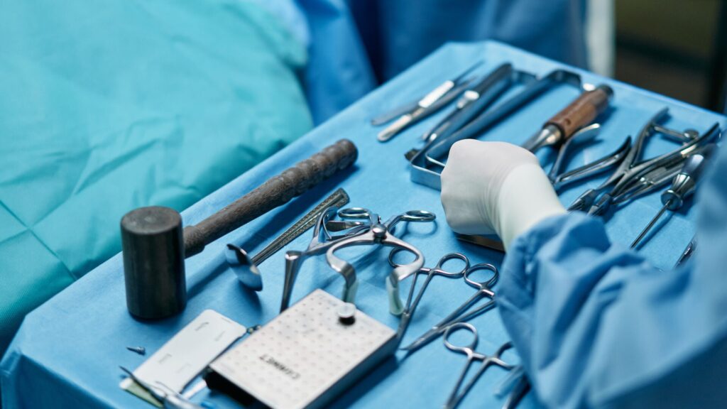
Use of Surgical Navigation for Craniopharyngioma Removal

Image sourced from Canva
The brain is the most complex and important part of the human body. It is in this area that all bodily functions are controlled, and it is responsible for making a person who they are. So, when it comes to caring for this part of the body, no expense must be spared, especially when abnormalities and ailments are found.
There are numerous ailments that can affect the brain, but this article will focus on craniopharyngioma and the intricate surgical navigation for craniopharyngioma removal.
What is Craniopharyngioma?
Craniopharyngioma is a rare tumor that grows near the hypothalamus and the pituitary gland, located at the base of the brain. This usually occurs in children 5 to 10 years old. However, note that it may also develop during adulthood.
There is no clear cause or origin for this. There are numerous studies that point out that this kind of tumor is developed in the early stages of pregnancy during embryogenesis, meaning one may develop the condition even before they are born.
This tumor is not considered cancerous, meaning it will not spread to other parts of the body but it can affect other bodily functions due to its sensitive location. The pituitary gland creates hormones, while the hypothalamus is responsible for releasing and controlling them. The danger then lies when the craniopharyngioma presses against these two structures in the brain.
All About Symptoms and Effects
Craniopharyngioma is not considered cancerous but symptoms and ailments may manifest when the tumor grows to reach a critical mass. As mentioned, it can increase cranial pressure and press on surrounding structures, thus affecting how one’s body works.
For example, it can create additional pressure in the brain, resulting in a condition called hydrocephalus. This can cause nausea, headache, vomiting, and issues with balance. If the tumor begins to press on the pituitary gland and hypothalamus, this can result in hormone imbalances and cause excessive thirst and urination, sleep disturbances, obesity, fatigue, decreased mental function, changes in personality and mood, and stunted growth and delayed puberty, especially for children.
Depending on its size and location, craniopharyngiomas can also affect one’s eyesight once the tumor begins to invade the optic nerve. If your child has an abrupt change in the quality of eyesight and exhibits slow growth accompanied by the symptoms stated above, it may be caused by craniopharyngioma.
Approaches and Modern Science
Craniopharyngiomas are usually diagnosed through various examinations and testing (physical, neurological, blood chemistry, and visual field exams). Scans and digital imaging are used to create a more accurate picture of what’s going on inside the brain.
This kind of tumor can be removed by surgery. Nowadays, minimally invasive neurosurgery is available and a full craniotomy is no longer required in most cases. It is possible to fully resect a craniopharyngioma, however, there are some cases where it can only be removed partially. In cases like this, surgery is partnered with other methods like radiation therapy.
There are two surgical approaches when it comes to removing craniopharyngiomas, namely transsphenoidal and transcranial.
The transsphenoidal approach is done by entering the brain passing through the bottom of the nostrils and sinuses or through an incision created under the lip. This remains to be the standard technique for cases involving the pituitary gland. The transcranial approach involves creating small incisions on the side of the head or the forehead to access the pituitary area.
Resection of craniopharyngiomas is considered to benefit from computer-assisted surgery. Because of the location of the craniopharyngioma near the brain and the skull base, a surgical navigation system might be used to verify the position of surgical tools during the operation.
Surgical navigation makes use of digital imaging, sensors, and robotics to give surgeons a clearer picture of what precisely is going on in the patient’s body before, during, and after surgery. This makes for fewer errors, shorter and less invasive surgical procedures, and faster recovery times.
Making Full Recovery Possible
It is important to consult only the best when it comes to issues with the brain. Removing craniopharyngiomas require a whole team of doctors and state-of-the-art facilities and equipment to properly tap into surgical navigation for craniopharyngioma removal.
At Minimally Invasive Neurosurgery of Texas (MINT), cutting-edge equipment, the Globus Excelsius Robotic Navigation, is used to provide only the best treatment. However, keep in mind that recovery depends on a lot of factors like the scope of tumor removal and the growth’s size.
Full recovery is possible but as with any medical procedure, there are risks involved. So, it is vital to choose a treatment option that is right for your condition—and of course, getting help from only the most credible neurosurgeons is just as important.
