
What Is Cervical Stenosis and Myelopathy Surgery in Lewisville TX?
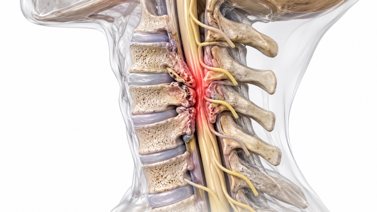
Cervical spinal stenosis is a narrowing of the spinal canal in the neck that can compress the spinal cord. When this compression produces neurological symptoms affecting the hands, arms, legs, balance, or bladder function, the condition is called cervical myelopathy. The medical community also refers to this condition as degenerative cervical myelopathy (DCM) or cervical spondylotic myelopathy (CSM).
Cervical myelopathy is the most common cause of acquired spinal cord dysfunction in adults and is more serious than cervical radiculopathy (a pinched nerve in the neck) because it involves the spinal cord itself rather than individual nerve roots. The spinal cord carries all motor and sensory signals between the brain and the body, so compression at the cervical level can affect function in the hands, arms, trunk, legs, bladder, and bowel.
Scott C. Kutz, MD, a board-certified myelopathy spine surgeon Lewisville TX, at Minimally Invasive Neurosurgery of Texas, evaluates and treats cervical stenosis and myelopathy at the practice’s Lewisville and Plano, Texas locations. Dr. Kutz is a Fellow of the American Association of Neurological Surgeons and the American College of Surgeons, with 27 years of neurosurgical experience. International guidelines consistently recommend surgery as the optimal treatment for patients with moderate to severe cervical myelopathy to prevent irreversible spinal cord damage (EFORT Open Reviews, 2025).
What Causes Cervical Stenosis and Myelopathy?
Cervical stenosis develops from age-related degenerative changes that gradually narrow the spinal canal. These changes include disc degeneration and bulging, bone spur (osteophyte) formation along the vertebral edges, thickening and buckling of the ligamentum flavum, facet joint enlargement from arthritis, and in some cases, ossification of the posterior longitudinal ligament (OPLL). These changes accumulate over time and progressively reduce the space available for the spinal cord.
Some patients are born with a naturally narrower cervical spinal canal, which means even minor degenerative changes can produce compression sooner. Others develop stenosis only after decades of progressive degeneration. The combination of a congenitally narrow canal and degenerative changes creates the highest risk for myelopathy.
Recognizing the Symptoms of Cervical Myelopathy
Cervical myelopathy symptoms develop gradually and are often subtle early on, making early recognition important. Common symptoms include difficulty with hand coordination and fine motor tasks such as buttoning a shirt, using utensils, or writing. Many patients notice they drop objects more frequently or struggle with tasks that require dexterity.
Gait instability and balance problems are another hallmark symptom. Patients may describe feeling unsteady, walking as if on a boat, or needing to hold onto walls and furniture for support. Numbness or tingling in the hands and feet, leg stiffness or heaviness, neck stiffness, and urinary urgency or frequency can also develop.
In advanced cases, patients may experience significant weakness in the arms or legs, loss of bladder or bowel control, and difficulty walking independently. These severe symptoms represent a medical urgency because they indicate substantial spinal cord compression that may cause permanent damage if not addressed.
Why Is Cervical Myelopathy Urgent?
Unlike many spinal conditions where patients can take time to explore conservative options, cervical myelopathy carries a risk of permanent, irreversible neurological damage if left untreated. The spinal cord has limited ability to recover once it is damaged by sustained compression. Research published in StatPearls (NIH, updated August 2025) notes that the prognosis worsens considerably if symptoms persist beyond 18 months without intervention.
This urgency does not mean every patient needs immediate surgery. Patients with mild symptoms and no evidence of spinal cord signal changes on MRI may be monitored closely with regular neurological examinations. However, progressive symptoms, significant spinal cord compression on imaging, T2 signal changes within the spinal cord on MRI, or any functional decline should prompt surgical evaluation without unnecessary delay.
The goal of surgery for myelopathy is to halt progression and preserve the neurological function that remains. In many cases, patients also experience meaningful improvement in symptoms after decompression. However, the degree of recovery depends on how long the compression has been present and how severe the damage has become before treatment.
Surgical Treatment Options for Cervical Myelopathy
The choice of surgical approach depends on where the compression occurs (front, back, or both), how many levels are involved, the patient’s cervical alignment, and the overall condition of the cervical spine.
Anterior cervical discectomy and fusion (ACDF) removes the compressing disc and bone spurs from the front of the neck and fuses the affected vertebrae. This is the most common approach for one- or two-level anterior compression causing myelopathy.
Cervical disc replacement may be appropriate for select myelopathy cases where compression is primarily from an anterior disc herniation at one or two levels, and the patient meets specific candidacy criteria. This motion-preserving option avoids fusion when feasible.
Anterior cervical corpectomy and fusion (ACCF) removes one or more vertebral bodies along with the adjacent discs to decompress the spinal cord from the front. This approach is used when the compression spans the height of the vertebral body rather than being limited to the disc level.
Posterior cervical laminectomy with fusion removes the lamina from the back of multiple vertebrae to decompress the spinal cord from behind, combined with instrumented fusion using screws and rods to maintain stability. This approach suits multilevel posterior compression.
Posterior cervical laminoplasty Lewisville TX creates a hinge in the lamina to expand the spinal canal without removing the bone entirely. The canal opens like a door, creating more space for the spinal cord while preserving some of the posterior structural elements. This approach avoids fusion and preserves more cervical motion than laminectomy with fusion.
Minimally invasive posterior cervical approaches, including cervical laminoforaminotomy, can address focal posterior compression at specific levels with reduced muscle disruption.
Dr. Kutz selects the approach based on the location, extent, and pattern of compression; the number of levels involved; cervical alignment; bone quality; and the patient’s overall health.
Who May Be a Candidate for Surgery?
Candidates for cervical stenosis or myelopathy surgery typically include patients with moderate to severe myelopathy symptoms such as gait difficulty, hand clumsiness, and balance problems. Patients with progressive neurological decline, even if currently mild, should be evaluated for surgery because waiting risks permanent damage. Patients with MRI evidence of spinal cord compression, especially those with T2 signal changes within the cord, are strong surgical candidates. Patients with cervical radiculopathy causing arm pain, numbness, or weakness alongside stenosis may also benefit from surgical decompression when conservative treatment has been inadequate.
Candidacy requires thorough neurological evaluation and imaging review. Contact Minimally Invasive Neurosurgery of Texas at (972) 244-3491.
Who May Not Be a Candidate?
Patients with mild, stable myelopathy symptoms and no MRI evidence of significant cord compression or signal changes may be managed conservatively with close monitoring and serial neurological examinations. Patients whose symptoms are caused by conditions other than cervical stenosis, such as peripheral neuropathy, vitamin B12 deficiency, multiple sclerosis, or normal pressure hydrocephalus, require different treatment. Patients with medical conditions that make surgery unsafe need an individual risk-benefit assessment that weighs the risks of surgery against the risks of progressive spinal cord damage.
How Is Cervical Myelopathy Surgery Performed?
The specific surgical steps vary by approach, but all cervical myelopathy procedures share the same fundamental goal: removing the tissue compressing the spinal cord to halt neurological deterioration and allow recovery.
Anterior approach (ACDF or ACCF). The patient lies face-up under general anesthesia. The surgeon makes a small incision at the front of the neck and gently retracts the soft tissues to access the cervical spine. The damaged disc (ACDF) or vertebral body (ACCF) is removed along with any bone spurs compressing the spinal cord. A cage or structural graft restores height, and a plate with screws stabilizes the segment. Fluoroscopy confirms proper positioning throughout.
Posterior approach (laminectomy with fusion or laminoplasty). The patient lies face-down under general anesthesia. The surgeon makes a midline incision at the back of the neck. For laminectomy with fusion, the lamina is removed at each affected level to open the spinal canal, and screws and rods are placed to stabilize the spine. For laminoplasty, a hinge is created on one side of the lamina, and the bone is lifted open on the other side and held in place with small plates. The spinal cord can then drift backward into the expanded canal, relieving the compression.
Dr. Kutz uses intraoperative neurophysiological monitoring during myelopathy procedures to continuously assess spinal cord function throughout the surgery.
Potential Benefits of Cervical Myelopathy Surgery
Surgical decompression for cervical myelopathy can halt the progression of spinal cord damage, improve gait and balance, improve hand coordination and dexterity, relieve neck and arm pain, restore or improve bladder function in some cases, and prevent the condition from progressing to severe disability or paralysis.
A prospective study of 278 patients with cervical spondylotic myelopathy reported a 2.5- to 3.5-point improvement in mJOA scores after surgery, regardless of approach, with significant quality-of-life improvements (Fehlings et al.). Patients with mild myelopathy who undergo early surgical intervention generally achieve the best functional outcomes. Patients with severe, long-standing myelopathy may stabilize rather than improve, which underscores the importance of early evaluation and treatment.
Risks and Complications
General surgical risks include infection, bleeding, blood clots, and adverse reactions to anesthesia. All spine surgery carries a small risk of nerve injury or spinal cord injury. C5 palsy is a specific complication that can occur after posterior cervical procedures, causing temporary deltoid weakness and difficulty raising the arm. This typically resolves over weeks to months. Dysphagia and hoarseness may occur temporarily after anterior cervical procedures due to retraction of the esophagus and proximity of the recurrent laryngeal nerve. Hardware failure, including screw loosening or plate displacement, is uncommon but possible. Pseudoarthrosis (failure of the fusion to heal) may cause persistent pain and instability. Adjacent segment disease can develop at levels above or below a fusion over time. Incomplete neurological recovery is a realistic possibility, particularly in patients with severe or long-standing myelopathy.
Recovery After Cervical Myelopathy Surgery
Recovery varies based on the severity of myelopathy before surgery, the specific procedure performed, and the number of levels treated. Hospital stays range from one to three days depending on the procedure and the patient’s overall condition. Walking begins the day of or after surgery. A cervical collar may be recommended for comfort and support during the initial healing period.
Physical and occupational therapy may be recommended to rebuild coordination, strength, grip, and balance. Return to desk work typically occurs within two to six weeks depending on the procedure. Physically demanding work requires three to six months of recovery. Neurological improvement may continue gradually for six to twelve months or longer after surgery as the spinal cord recovers from compression.
Patients with mild myelopathy who undergo early surgery generally recover the most function. Patients with severe myelopathy may stabilize rather than improve dramatically, which is still meaningful because it prevents further decline.
Alternatives to Surgery
Conservative monitoring with regular neurological examinations may be appropriate for patients with mild, stable symptoms and no evidence of progressive cord compression. Conservative treatment cannot reverse spinal cord compression, but monitoring ensures that surgery can be offered promptly if progression occurs.
Physical therapy can improve neck flexibility, posture, and core stability, which may help manage symptoms but does not address the underlying structural compression.
Cervical epidural injections may provide temporary relief of radicular arm pain associated with stenosis but do not treat spinal cord compression.
Activity modification such as avoiding high-risk activities (contact sports, heavy overhead work) can reduce the risk of acute spinal cord injury in patients with significant stenosis who are not yet surgical candidates.
Frequently Asked Questions
What is cervical myelopathy?
Cervical myelopathy is compression of the spinal cord in the neck caused by narrowing of the spinal canal. It produces symptoms including hand clumsiness, difficulty with fine motor tasks, gait instability, balance problems, and numbness in the hands and feet. Without treatment, it can progress to permanent neurological damage.
Is cervical myelopathy serious?
Yes. Cervical myelopathy involves the spinal cord, which has limited ability to recover once damaged. International guidelines recommend surgery for moderate to severe cases to halt neurological decline and prevent irreversible disability.
What warning signs should prompt me to see a spine surgeon?
Warning signs include difficulty with hand coordination, dropping objects, changes in handwriting, unsteady walking, frequent stumbling, progressive numbness in the hands and feet, and neck stiffness that worsens over time. Bladder or bowel changes require urgent evaluation.
Can cervical myelopathy get better without surgery?
Mild cases may stabilize, but conservative treatment cannot reverse spinal cord compression or restore function that has already been lost. Research shows that symptoms lasting beyond 18 months without treatment are associated with significantly worse outcomes.
What surgery treats cervical myelopathy?
Options include anterior cervical discectomy and fusion (ACDF), cervical disc replacement, anterior corpectomy and fusion (ACCF), posterior laminectomy with fusion, and posterior laminoplasty. The choice depends on where the compression occurs and how many levels are involved.
How long does recovery take after cervical myelopathy surgery?
Hospital stays are typically one to three days. Return to desk work occurs within two to six weeks. Physical and occupational therapy supports recovery of coordination and balance. Neurological improvement may continue for six to twelve months or longer.
What is the difference between cervical stenosis and cervical myelopathy?
Cervical stenosis is the anatomical narrowing of the spinal canal. Cervical myelopathy is the clinical syndrome that develops when that narrowing compresses the spinal cord and causes neurological symptoms. A patient can have stenosis on MRI without myelopathy symptoms.
What is a C5 palsy?
C5 palsy is a specific complication that can occur after posterior cervical surgery, causing temporary weakness in the deltoid muscle and difficulty raising the arm at the shoulder. It typically resolves over weeks to months without additional surgery.
Who treats cervical stenosis and myelopathy in Lewisville and Plano TX?
Scott C. Kutz, MD, at Minimally Invasive Neurosurgery of Texas. Call (972) 244-3491 to schedule an evaluation.
Schedule a Consultation for Cervical Stenosis and Myelopathy
Scott C. Kutz, MD, is a board-certified neurosurgeon serving patients at Minimally Invasive Neurosurgery of Texas in Lewisville and Plano, Texas. To discuss whether cervical stenosis and myelopathy is appropriate for your condition, call (972) 244-3491 or request an appointment online.
- Lewisville: 1850 Lakepointe Drive, Suite 500, Lewisville, TX 75057
- Plano: 5465 Legacy Drive, Suite 650, Plano, TX 75024