
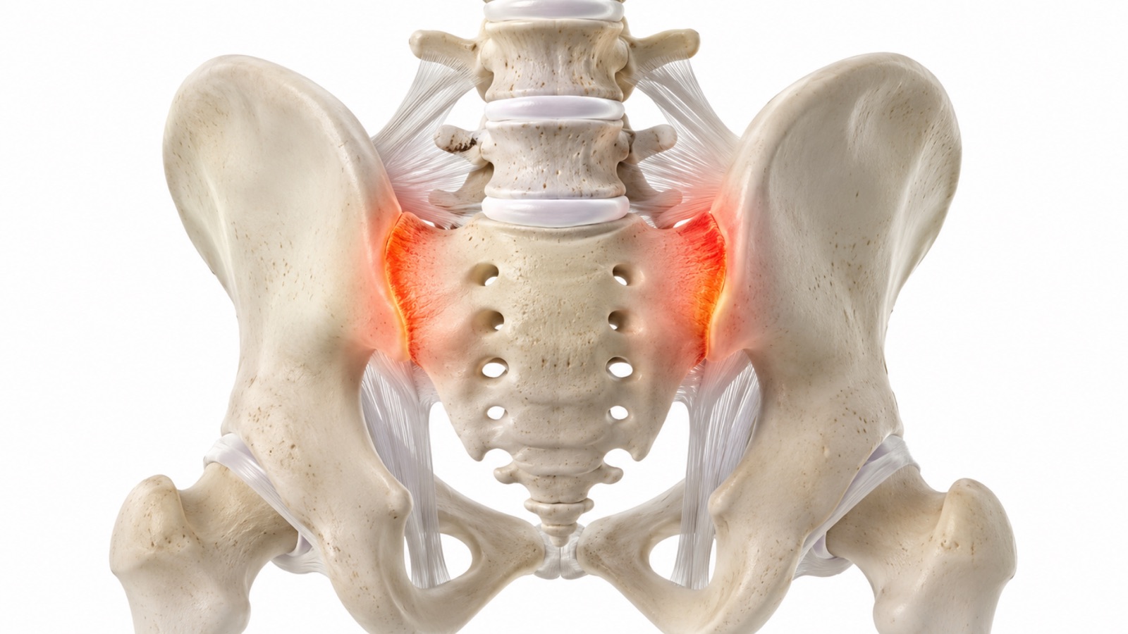
What Is SI Joint Fusion Surgery in Lewisville TX?
The sacroiliac (SI) joints connect the sacrum (the triangular bone at the base of the spine) to the iliac bones of the pelvis. These two joints sit on either side of the lower spine and serve as the structural bridge between the spine and the lower body. The SI joints transmit the weight of the upper body to the pelvis and legs, and they absorb shock during walking, running, and bending. Unlike most joints in the body, the SI joints are designed for stability rather than large ranges of motion. They move only a few degrees in any direction.
When an SI joint becomes dysfunctional due to degeneration, inflammation, injury, or altered mechanics after lumbar fusion, it can produce chronic pain in the lower back, buttock, hip, and groin that significantly limits daily activities. SI joint dysfunction is a commonly underdiagnosed cause of lower back pain. Studies suggest that the SI joint is the primary pain source in approximately 15 to 30 percent of patients presenting with chronic lower back pain.
SI joint fusion is a minimally invasive surgical procedure that stabilizes the dysfunctional joint by placing implants across it to promote bony fusion and eliminate the painful motion. Scott C. Kutz, MD, a board-certified neurosurgeon at Minimally Invasive Neurosurgery of Texas, performs minimally invasive SI joint fusion at the practice’s Lewisville and Plano, Texas locations. Dr. Kutz has published peer-reviewed content on sacroiliac joint fusion through Spine-Health, including articles on what patients should know about the procedure, how it is performed, and what to expect during recovery.
What Conditions Does SI Joint Surgery Treat?
Sacroiliac joint dysfunction is the primary indication for SI joint fusion. This happens when the SI joint becomes a chronic pain source because of degeneration, hypermobility, or inflammation. Pain is typically felt in the lower back and buttock on one side, and it often worsens with prolonged sitting, standing from a seated position, climbing stairs, or rolling over in bed.
SI joint pain after lumbar fusion is a recognized complication of lumbar spinal fusion. When the lumbar spine is fused, the segments that previously shared the workload of movement and load transfer no longer move. This altered biomechanics transfers additional stress to the SI joints, causing them to become symptomatic. The incidence of SI joint pain following lumbar fusion has been reported at up to 40 percent in some studies.
Sacroiliitis is inflammation of the SI joint that may result from mechanical stress, degenerative changes, or inflammatory conditions. When sacroiliitis persists despite conservative treatment, SI joint fusion may be considered to eliminate the painful motion and inflammatory cycle.
SI joint instability from trauma (such as a fall or motor vehicle accident), pregnancy-related ligamentous laxity that does not resolve postpartum, or degenerative weakening of the joint ligaments can cause the joint to move excessively, producing chronic pain with weight-bearing activities.
Degenerative sacroiliitis occurs as the joint surfaces wear down over time, producing bone-on-bone contact, osteophyte formation, and chronic inflammation. This is more common in older adults and in patients who have undergone prior lumbar fusion.
Who May Be a Candidate for SI Joint Surgery?
Candidates for SI joint fusion have chronic SI joint pain that meets specific diagnostic and treatment criteria.
Duration of symptoms. Pain should have been present for at least six months and should be significantly limiting daily function, work, or quality of life.
Failed conservative treatment. Patients should have completed an adequate course of conservative care including physical therapy focused on core stabilization and pelvic mechanics, anti-inflammatory medications, activity modification, and at least one course of SI joint injections.
Confirmed diagnosis through diagnostic SI joint injection. The diagnosis of SI joint dysfunction is confirmed when injecting numbing medication directly into the SI joint under fluoroscopic guidance temporarily eliminates or substantially reduces the patient’s pain. This diagnostic injection is the most important step in establishing that the SI joint is the true pain source, not a bystander. Without a positive diagnostic injection, SI joint fusion should not be performed.
Imaging findings. While imaging is not always diagnostic for SI joint dysfunction, CT or MRI may show degenerative changes at the SI joint, sclerosis, joint space narrowing, or osteophytes that support the clinical diagnosis.
Contact Minimally Invasive Neurosurgery of Texas at (972) 244-3491.
Who May Not Be a Candidate?
Patients whose pain has not been confirmed by diagnostic SI joint injections. Without objective confirmation that the SI joint is the source of pain, proceeding with fusion risks operating on the wrong structure.
Patients with active infection at the surgical site or systemically are not candidates until the infection is fully treated.
Patients with inflammatory arthropathy (such as ankylosing spondylitis or psoriatic arthritis affecting the SI joints) that is better managed with medical therapy. These conditions may respond to biologic medications and should be co-managed with a rheumatologist.
Patients who have not completed adequate conservative treatment. Physical therapy, injections, and medication management should be given a reasonable trial before surgery is considered.
Patients with significant untreated hip pathology. Hip joint problems can mimic or coexist with SI joint pain. If hip pathology contributes to the symptoms, address it independently or concurrently.
Patients whose pain pattern does not match the SI joint distribution. SI joint pain follows a characteristic pattern centered in the lower back and buttock, sometimes extending into the posterior thigh. Pain that does not follow this distribution may originate from a different structure.
How Is Minimally Invasive SI Joint Fusion Performed?
Minimally invasive SI joint fusion is performed through a small incision, typically two to three centimeters, on the side of the buttock. The patient is positioned face down on the operating table under general anesthesia.
Using fluoroscopic guidance (real-time X-ray imaging), the surgeon identifies the SI joint and creates a channel through the iliac bone into the sacrum, crossing the joint. The surgeon places two to three small implants across the SI joint through this channel. The implants serve two purposes: they immediately stabilize the joint, reducing painful motion, and they promote bony fusion over time by encouraging bone to grow across and through the implant surfaces.
The procedure typically takes 45 to 60 minutes. The small incision and targeted approach spare the surrounding muscles and ligaments. No tendons are detached, and no significant muscle dissection is required.
Most patients go home the same day or after one night of observation. The procedure fits within outpatient spine surgery protocols for appropriate candidates.
Dr. Kutz has published detailed information about how SI joint fusion is performed, what patients should know before the procedure, and what to expect during recovery through his Spine-Health author profile.
Potential Benefits
Minimally invasive SI joint fusion offers several advantages for patients with confirmed SI joint dysfunction who have not responded to conservative care.
Targeted pain relief. When diagnostic injections confirm the SI joint as the pain source, fusion directly addresses the problem by eliminating painful motion at that joint.
Small incision with minimal tissue disruption. The lateral approach uses a two-to-three-centimeter incision and does not require detachment of muscles or tendons, resulting in less post-operative pain and faster wound healing.
Short operative time. The procedure typically takes less than one hour, reducing time under general anesthesia and the associated physiological stress.
Same-day or next-day discharge. Most patients leave the surgical facility within hours of the procedure or after a single overnight stay.
Structured recovery with progressive return to activity. Patients follow a predictable recovery timeline with clear milestones for resuming daily activities, work, and exercise.
Published clinical evidence. Peer-reviewed studies demonstrate significant pain reduction and functional improvement in patients who meet proper selection criteria for minimally invasive SI joint fusion.
Alternatives to SI Joint Surgery
SI joint dysfunction does not always require surgery. Many patients achieve adequate relief through nonsurgical approaches, and Dr. Kutz evaluates all options before recommending fusion.
Physical therapy focused on core stabilization, pelvic floor strengthening, hip mechanics, and postural correction is the foundation of conservative management. A therapist experienced with SI joint dysfunction can teach specific exercises and manual techniques to reduce joint irritation.
SI joint injections using corticosteroid and local anesthetic can provide temporary to lasting pain relief. Some patients achieve months of meaningful improvement from periodic injections and may not need surgery.
Radiofrequency ablation of the nerves supplying the SI joint can reduce pain signals for six to twelve months or longer. This procedure burns the small sensory nerves around the joint, blocking pain transmission without altering the joint itself.
Oral medications including anti-inflammatory drugs, acetaminophen, and short-term use of muscle relaxants may manage symptoms during acute flares.
SI joint belts and bracing can provide external stabilization that reduces joint motion and pain during weight-bearing activities, particularly during acute episodes.
Activity modification, including avoiding prolonged sitting, using proper body mechanics during lifting, and adjusting sleep positions, can reduce SI joint irritation.
Dr. Kutz recommends fusion only after conservative measures have been adequately tried and diagnostic injection has confirmed the SI joint as the pain source.
Risks and Complications
As with any surgical procedure, SI joint fusion carries risks.
General surgical risks include infection, bleeding, blood clots, and adverse reactions to anesthesia. These are uncommon with minimally invasive SI joint fusion due to the small incision and short operative time.
Nerve irritation may occur if an implant is positioned near a sacral nerve root. Careful fluoroscopic guidance during implant placement minimizes this risk.
Implant malposition is possible but uncommon with real-time imaging guidance. A malpositioned implant may require adjustment or removal.
Failure to fuse can occur if the bone does not grow across the joint as expected. Risk factors for nonunion include smoking, diabetes, osteoporosis, and noncompliance with post-operative weight-bearing restrictions.
Persistent pain may continue after fusion if additional pain generators exist beyond the SI joint, or if the diagnosis was not adequately confirmed before surgery.
Need for additional surgery is possible if the fusion does not heal, if an implant requires revision, or if the contralateral (opposite side) SI joint becomes symptomatic over time.
Dr. Kutz discusses all relevant risks with each patient and uses proper diagnostic confirmation before recommending surgery to maximize the likelihood of a successful outcome.
Recovery
First two weeks. Most patients go home the same day or after one night. Crutches or a walker are recommended for two to four weeks to limit weight-bearing on the surgical side and protect the implants while early healing occurs. Ice therapy and prescribed medications manage post-operative discomfort. Short, frequent walks are encouraged from day one.
Weeks two through six. Sitting tolerance improves gradually. Most patients return to desk work within two to four weeks. Driving is typically safe once the patient is off narcotic medications and can comfortably operate the vehicle, usually within two to three weeks. Physical therapy may begin during this period, focusing on gentle range of motion and core activation.
Months two through six. Progressive increase in activity. Physical therapy advances to include core strengthening, hip strengthening, and pelvic stability exercises. The fusion matures over this period as bone grows across the joint.
Full activity clearance usually occurs at three to six months, once imaging confirms adequate fusion progress. Impact activities and heavy lifting are restricted until the surgeon confirms sufficient healing.
When to seek immediate medical attention. Contact the office or go to the emergency room if you experience sudden or progressive weakness in your legs, loss of bladder or bowel control, high fever, or worsening pain that is not controlled by your prescribed medications.
Why Choose Minimally Invasive Neurosurgery of Texas for SI Joint Surgery?
SI joint dysfunction is frequently misdiagnosed as lumbar spine pathology, hip pathology, or generic “lower back pain.” Accurate diagnosis requires a methodical approach and a willingness to look beyond the lumbar spine when lumbar imaging does not explain the patient’s symptoms. Dr. Kutz brings 27 years of neurosurgical experience and a published track record on SI joint fusion through Spine-Health to every evaluation.
Dr. Kutz has authored peer-reviewed content on sacroiliac joint fusion, including articles on what patients should know about the procedure, how it is performed, and what to expect during recovery. This published expertise reflects his depth of experience with SI joint pathology.
Minimally Invasive Neurosurgery of Texas serves patients at two North Texas locations:
Lewisville office: 1850 Lakepointe Drive, Suite 500, Lewisville, TX 75057 Plano office: 5465 Legacy Drive, Suite 650, Plano, TX 75024
Patients seeking SI joint fusion surgery Lewisville TX can schedule a consultation by calling (972) 244-3491 or visit the contact page.
Frequently Asked Questions
What is the sacroiliac joint?
The SI joints connect the base of the spine (sacrum) to the pelvis (iliac bones). There is one on each side. They transmit upper body weight to the lower body and absorb shock during movement. Unlike the hip or knee, the SI joints are designed for stability and move only a few degrees.
How do you know the SI joint is causing my pain?
We confirm the diagnosis with diagnostic SI joint injections. A physician injects numbing medication directly into the SI joint under fluoroscopic guidance. If this injection temporarily eliminates or substantially reduces the pain, the SI joint is confirmed as the source. This step is required before recommending SI joint fusion.
What does SI joint pain feel like?
SI joint pain is typically felt in the lower back and buttock on one side. It may extend into the back of the thigh or groin. Pain often worsens with prolonged sitting, standing from a seated position, climbing stairs, walking uphill, or rolling over in bed. People often mistake it for lumbar spine or hip problems.
Is SI joint fusion minimally invasive?
Yes. The procedure uses a small incision (two to three centimeters) on the side of the buttock. The surgeon places two to three implants across the joint under fluoroscopic guidance. Most patients go home the same day or after one night.
How long does recovery take after SI joint fusion?
Crutches or a walker for two to four weeks. Return to desk work typically within two to four weeks. Progressive increase in activity over months two through six. Full activity clearance at three to six months once fusion is confirmed on imaging.
Can SI joint pain develop after lumbar fusion?
Yes. Altered biomechanics from a lumbar fusion can transfer additional stress to the SI joints, causing them to become symptomatic. Some studies report this complication in up to 40 percent of lumbar fusion patients.
What happens if I do not have SI joint surgery?
Nonsurgical options include physical therapy, SI joint injections, radiofrequency ablation, bracing, and medication management. Many patients manage their symptoms effectively without surgery. Fusion is recommended only when conservative measures have failed, and the SI joint has been confirmed as the pain source.
Are both SI joints treated at the same time?
Typically, providers treat only the symptomatic joint. If separate diagnostic injections confirm both joints as pain sources, providers may address the second joint in a separate procedure after recovery from the first.
Who performs SI joint fusion in Lewisville and Plano TX?
Scott C. Kutz, MD, at Minimally Invasive Neurosurgery of Texas. Dr. Kutz has published peer-reviewed content on SI joint fusion through Spine-Health. Call (972) 244-3491.
Schedule a Consultation for Sacroiliac Joint Surgery
Scott C. Kutz, MD, is a board-certified neurosurgeon serving patients at Minimally Invasive Neurosurgery of Texas in Lewisville and Plano, Texas. To discuss whether sacroiliac joint surgery is appropriate for your condition, call (972) 244-3491 or request an appointment online.
- Lewisville: 1850 Lakepointe Drive, Suite 500, Lewisville, TX 75057
- Plano: 5465 Legacy Drive, Suite 650, Plano, TX 75024