
What Is Minimally Invasive Spine Surgery in Lewisville TX?
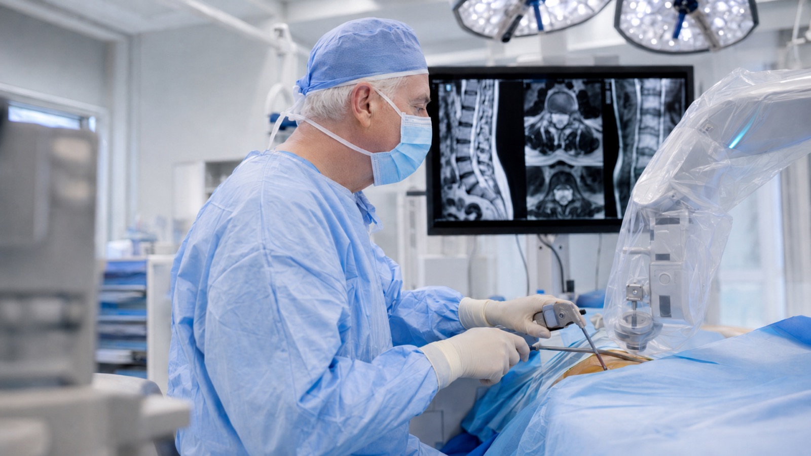
Minimally invasive spine surgery is a category of surgical techniques that treat spinal conditions through small incisions, typically one inch or less, instead of the large open incisions used in traditional spine surgery. The surgeon accesses the spine using tubular retractors, endoscopes, or specialized instruments that dilate and separate muscle fibers rather than cutting through them. This approach reduces damage to the muscles, ligaments, and soft tissues surrounding the spine, which directly translates to less post-operative pain, shorter hospital stays, and faster recovery for patients.
Scott C. Kutz, MD, a board-certified neurosurgeon at Minimally Invasive Neurosurgery of Texas, has performed over 6,000 successful spine surgeries using minimally invasive techniques at the practice’s Lewisville and Plano, Texas locations. Dr. Kutz is a Fellow of the American Association of Neurological Surgeons and the American College of Surgeons, with 27 years of neurosurgical experience. The practice specializes in muscle-sparing, motion-preserving approaches that achieve the same surgical goals as open surgery while reducing the physical toll on the patient’s body.
A 2025 review published in Neurosurgery confirmed that minimally invasive spine surgery has transformed spinal care by minimizing tissue disruption, reducing recovery times, and lowering complication rates compared with traditional open procedures (Ikwuegbuenyi et al., Neurosurgery, 2025). The clinical outcomes for decompression and fusion procedures performed minimally invasively are comparable to open surgery, with the added benefits of a less traumatic surgical experience.
How Does Minimally Invasive Spine Surgery Differ from Open Surgery?
Traditional open spine surgery requires an incision of three to six inches or longer. The surgeon must strip the paraspinal muscles away from the spine to see and access the vertebrae, discs, and nerves. This muscle detachment is a major source of post-operative pain and a primary reason recovery from open surgery takes weeks to months. Significant blood loss is common, and the larger wound carries a higher risk of infection.
Minimally invasive spine surgery achieves the same surgical objectives through a fundamentally different access strategy. Instead of cutting through muscle, the surgeon uses a series of progressively larger dilators to gently push muscle fibers apart and create a narrow working channel to the spine. A tubular retractor holds this channel open while the surgeon works through it using specialized instruments, an operating microscope, or an endoscope for visualization. Real-time imaging guidance, including fluoroscopy (live X-ray) and, in some cases, the Globus Excelsius GPS Robotic Navigation Platform or Augmedics augmented reality system used at Minimally Invasive Neurosurgery of Texas, provides precise three-dimensional guidance throughout the procedure.
The practical impact for patients is significant. Smaller incisions heal faster. Preserved muscles recover their function sooner. Reduced blood loss decreases the chance of needing a transfusion. Many minimally invasive procedures can be performed on an outpatient basis or with a single overnight stay, compared to multi-day hospitalizations for open surgery. These advantages do not come at the cost of surgical effectiveness. When performed by an experienced surgeon, minimally invasive decompression, stabilization, and fusion produce long-term outcomes equivalent to open approaches.
What Conditions Can Be Treated with Minimally Invasive Spine Surgery?
Minimally invasive techniques can address the majority of common spinal conditions that require surgical intervention. The specific technique Dr. Kutz selects depends on the diagnosis, the location within the spine, and the goals of surgery.
Minimally invasive discectomy can treat herniated discs in the cervical, thoracic, or lumbar spine. The surgeon removes the portion of the disc pressing on the nerve while preserving the remainder of the disc and surrounding structures.
Spinal stenosis and lumbar spinal stenosis are treated through minimally invasive laminectomy or laminotomy, which removes the bone and thickened ligament compressing the spinal cord or nerve roots.
Degenerative disc disease that causes instability or chronic pain unresponsive to conservative care may require fusion or artificial disc replacement, both of which can be performed through minimally invasive approaches.
Spondylolisthesis occurs when one vertebra slips forward over the one below it. Minimally invasive fusion techniques stabilize the affected segment and relieve nerve compression.
Sciatica and lumbar radiculopathy caused by disc herniation or foraminal narrowing often respond well to minimally invasive decompression.
Sacroiliac joint dysfunction can be treated with minimally invasive SI joint fusion when conservative measures have failed.
Vertebral compression fractures from osteoporosis or trauma may be stabilized through minimally invasive vertebroplasty or kyphoplasty.
Spinal tumors and infections can be biopsied or treated through minimally invasive approaches in appropriate cases.
What Minimally Invasive Techniques Does MINT Perform?
Dr. Kutz performs a comprehensive range of minimally invasive spine techniques at Minimally Invasive Neurosurgery of Texas. Each technique targets a specific spinal problem and region.
Cervical laminoforaminotomy widens the bony canal where a nerve root exits the cervical spine, relieving pressure caused by bone spurs or disc fragments in the neck.
Posterior cervical facet fusion stabilizes the cervical spine through a posterior approach when instability or degeneration affects the facet joints.
Lumbar laminotomy and discectomy remove herniated disc material and, when necessary, a small amount of bone to decompress a pinched nerve in the lower back.
Lumbar foraminotomy and lateral discectomy open the neural foramen, the bony exit window on the side of the spine, to relieve nerve root compression.
Transforaminal lumbar interbody fusion (TLIF) approaches the disc space from the side through the neural foramen, allowing the surgeon to remove the damaged disc, insert a cage filled with bone graft material, and stabilize the segment with percutaneous screws, all through small incisions.
Lateral lumbar interbody fusion (LLIF) accesses the disc space through a small incision on the patient’s side, avoiding the back muscles entirely. This approach suits conditions involving disc collapse or instability at multiple levels.
Oblique lumbar interbody fusion (OLIF) approaches the spine at an oblique angle from the front side, providing access to the disc space while avoiding both the back muscles and the psoas muscle.
Percutaneous spinal instrumentation places pedicle screws through small stab incisions using fluoroscopic or robotic guidance, eliminating the need for a long open incision to expose the spine for screw placement.
Thoracic discectomy and interbody fusion treat conditions in the mid-back through small incisions, which is particularly valuable given the technical difficulty of accessing the thoracic spine.
Sacroiliac joint fusion stabilizes the SI joint through a small lateral incision when chronic SI joint dysfunction has not responded to conservative treatment.
Endoscopic spine surgery represents the least invasive approach in the MISS category, using a quarter-inch incision and a camera-equipped endoscope. Dr. Kutz holds Endoscopic Spine Surgery Level 1-2 Workshop certification.
Who May Be a Candidate for Minimally Invasive Spine Surgery?
Candidates for minimally invasive spine surgery are patients with a clearly diagnosed spinal condition that has not improved with appropriate nonsurgical treatment and whose anatomy allows safe access through a minimally invasive approach.
Symptoms that may indicate surgical candidacy include persistent or worsening pain in the back, neck, arms, or legs that interferes with daily activities and sleep. Numbness, tingling, or weakness in the arms or legs that corresponds to a specific nerve distribution suggests nerve compression. Difficulty walking, loss of balance, or a sensation of heaviness in the legs may indicate spinal cord or nerve root compression.
Conservative treatment should come first. Dr. Kutz typically recommends that patients try physical therapy, anti-inflammatory medications, and, when appropriate, guided injections before considering surgery. Surgery becomes a reasonable next step when these measures have not provided adequate relief after a sustained effort, typically six to twelve weeks or longer depending on the condition.
Imaging must confirm a structural cause. An MRI or CT scan should demonstrate a specific anatomical problem, such as a herniated disc, spinal stenosis, or spondylolisthesis, that correlates with the patient’s symptoms and physical examination findings. Imaging abnormalities without corresponding symptoms do not justify surgery alone.
Patient health factors. Minimally invasive approaches may be particularly beneficial for older patients, those with diabetes, those who are overweight, or those who have other medical conditions that increase the risk of complications from open surgery. The smaller incision, reduced blood loss, and shorter anesthesia time associated with MISS reduce the procedure’s overall physiological stress.
Candidacy requires an individualized evaluation. To schedule a consultation, contact Minimally Invasive Neurosurgery of Texas at (972) 244-3491.
Who May Not Be a Candidate?
Not every spinal condition or patient anatomy suits a minimally invasive approach. Recognizing these limitations helps set realistic expectations.
Severe multilevel spinal deformity. Conditions involving significant scoliosis, kyphosis, or complex deformity correction may require the broader surgical exposure that open surgery provides.
Certain revision surgeries. Patients who have had prior spine surgery with extensive hardware or significant scar tissue may need an open approach to navigate the altered anatomy safely. However, some revision cases can still be addressed minimally invasively.
Conditions requiring extensive decompression. When stenosis or compression involves many spinal levels, the surgeon may need the wider field of view and working space that a traditional approach provides.
Anatomical limitations. Some patients have bone structure, body habitus, or spinal anatomy that makes safe tubular or endoscopic access technically challenging.
Active spinal infection or tumor requiring wide excision. While minimally invasive techniques can be used for biopsy and some tumor procedures, conditions requiring aggressive resection with wide margins typically need open surgery.
The decision between minimally invasive and open surgery is not about choosing a “better” or “worse” option. It is about matching the surgical approach to the specific problem and the individual patient. Dr. Kutz evaluates each case and recommends the approach most likely to produce the best long-term result.
Potential Benefits of Minimally Invasive Spine Surgery
The benefits of minimally invasive techniques are well documented in peer-reviewed literature. While individual outcomes vary, the general advantages over traditional open surgery include:
Less muscle and soft tissue damage. By working between muscle fibers instead of cutting through or detaching them, MISS preserves the muscles that support the spine. This directly reduces post-operative pain and accelerates functional recovery.
Smaller incisions. Incisions typically range from half an inch to one inch, compared to three to six inches or more for open surgery. Smaller incisions heal faster, produce less scarring, and have a lower risk of wound complications and infection.
Reduced blood loss. Preserving the soft tissue envelope and working through a narrow channel significantly reduces intraoperative bleeding, lowering the chance of requiring a blood transfusion.
Shorter hospital stays. Many minimally invasive decompression procedures are performed on an outpatient basis. Minimally invasive fusions often require only a one-night hospital stay, compared to two to four days for open fusion.
Faster recovery. Patients undergoing minimally invasive procedures typically return to light activities and desk work within two to four weeks. Return to full activity usually occurs within six to twelve weeks, depending on the procedure and the patient’s condition.
Reduced narcotic use. Less tissue trauma means less post-surgical pain, which reduces the need for opioid pain medications and their associated side effects.
Comparable long-term outcomes. Research consistently demonstrates that minimally invasive techniques produce pain relief, functional improvement, and fusion rates equivalent to open surgery when performed by experienced surgeons.
These benefits represent what the clinical evidence generally demonstrates. No surgical procedure guarantees a specific outcome, and Dr. Kutz discusses realistic expectations with each patient during the pre-operative consultation.
Risks and Complications
All surgical procedures carry risk, including minimally invasive spine surgery. Patients should understand general and procedure-specific risks before deciding on treatment.
General surgical risks apply to any operation and include infection, bleeding, blood clots, and adverse reactions to anesthesia. These risks are generally lower with minimally invasive approaches due to the smaller incision and reduced tissue disruption, but they cannot be eliminated.
Nerve injury is a rare but serious possibility in any spine surgery. The surgeon works in proximity to the spinal cord and nerve roots. Advanced imaging guidance and magnification tools help minimize this risk.
Incomplete decompression may occur if the limited working field of a minimally invasive approach does not allow the surgeon to address the pathology fully. In rare cases, conversion to an open approach during the same operation may be necessary.
A dural tear can occur when the thin membrane surrounding the spinal cord and nerves is inadvertently punctured. This can cause a spinal fluid leak and headache. Most dural tears are repaired during the same procedure or heal on their own.
Hardware misplacement is a risk with any procedure involving screws or instrumentation. Robotic navigation technology, such as the Globus Excelsius GPS used at MINT, provides sub-millimeter guidance accuracy that significantly reduces this risk.
Pseudoarthrosis can occur after fusion procedures when the bone graft does not fully heal into a solid mass. This may cause persistent pain and potentially require additional surgery.
Recurrent herniation is possible after any discectomy procedure. Disc material can re-herniate at the same or a different level.
Adjacent segment disease may develop over time when the spinal levels above or below a fused segment experience accelerated wear due to altered biomechanics. Motion-preserving techniques, including artificial disc replacement, aim to reduce this risk.
Dr. Kutz discusses all risks relevant to the specific procedure with each patient and answers questions about how those risks apply to their individual situation.
Recovery After Minimally Invasive Spine Surgery
Recovery timelines vary based on the type of procedure performed, the condition treated, the patient’s age, overall health, and physical demands. The following are general guidelines.
Decompression procedures (discectomy, laminectomy, foraminotomy)
These procedures typically allow same-day discharge or a single overnight stay. Patients usually walk within hours of surgery. Return to desk work often occurs within one to two weeks. Return to physically demanding work usually requires four to six weeks. Light exercise and walking are encouraged early in recovery.
Fusion procedures (TLIF, LLIF, OLIF)
Fusion procedures generally require one to two nights in the hospital. Patients walk the day of or the day after surgery. Return to desk work typically occurs within two to four weeks. The bone graft needs time to mature and solidify, so heavy lifting and strenuous activity are restricted for three to six months depending on the number of levels fused. Follow-up imaging monitors the fusion progress.
General recovery guidelines for all procedures
The first week focuses on short, frequent walks and basic self-care. Pain is typically managed with a combination of over-the-counter medications and a short course of prescribed medication when needed. Ice therapy helps manage swelling at the incision site. Driving is usually safe once the patient is alert and off narcotic medications, typically within one to two weeks. Physical therapy may be recommended to rebuild strength and flexibility, depending on the procedure and the patient’s baseline condition.
When to seek immediate medical attention. Contact the office or go to the emergency room if you experience sudden or progressive weakness in your arms or legs, loss of bladder or bowel control, high fever with severe back pain, or worsening pain that is not controlled by your prescribed medications.
Dr. Kutz provides individualized recovery instructions and schedules follow-up visits based on each patient’s progress.
Technology at Minimally Invasive Neurosurgery of Texas
The quality and precision of minimally invasive spine surgery depend heavily on the technology available to the surgeon. Minimally Invasive Neurosurgery of Texas uses advanced systems that enhance accuracy, safety, and outcomes.
Globus Excelsius GPS Robotic Navigation Platform combines real-time imaging with a rigid robotic arm to guide instrument and screw placement with sub-millimeter accuracy. This technology is especially valuable for percutaneous screw placement, where the surgeon cannot directly see the bony anatomy. The robotic arm provides a steady, precise trajectory that reduces the risk of screw misplacement and lowers radiation exposure compared to conventional fluoroscopy-guided techniques.
Augmedics augmented reality system projects a three-dimensional image of the patient’s spine onto a transparent headset worn by the surgeon. This lets Dr. Kutz visualize internal spinal structures while looking directly at the patient, rather than turning away to view a separate monitor. The result is more intuitive navigation and improved ergonomics during complex procedures.
Endoscopic spine surgery uses a high-definition camera and specialized instruments through the smallest possible incision, as described on the dedicated endoscopic spine surgery page.
Intraoperative fluoroscopy provides continuous real-time X-ray imaging throughout the procedure, confirming instrument position and spinal alignment at every step.
These technologies do not replace surgical skill and judgment. They enhance an experienced surgeon’s capabilities, allowing Dr. Kutz to perform procedures with greater precision and confidence while minimizing risk to surrounding structures.
Why Choose Minimally Invasive Neurosurgery of Texas?
Minimally Invasive Neurosurgery of Texas was founded on the principle that spine surgery should be as precise, efficient, and gentle as modern technology allows. The practice name reflects this commitment. Every procedure Dr. Kutz performs prioritizes the least invasive approach that can effectively address the patient’s condition.
Dr. Kutz brings 27 years of neurosurgical experience, board certification from the American Board of Neurological Surgery, and fellowships in both the American Association of Neurological Surgeons and the American College of Surgeons. He has published peer-reviewed content on Spine-Health, including articles on sacroiliac joint fusion and how posture affects lumbar herniated discs. His training includes a neurosurgical residency at Louisiana State University and a fellowship in stereotactic and functional neurosurgery at Rush University Medical Center in Chicago.
The practice integrates robotic navigation, augmented reality, and endoscopic technology to provide patients with the most advanced minimally invasive options available. MINT’s surgical team takes time to understand each patient’s condition, explain treatment options, and develop a plan tailored to the individual’s anatomy, goals, and lifestyle. Patients receive direct, personal attention from their surgeon throughout evaluation, surgery, and recovery.
Minimally Invasive Neurosurgery of Texas serves patients at two North Texas locations:
Lewisville office: 1850 Lakepointe Drive, Suite 500, Lewisville, TX 75057 Plano office: 5465 Legacy Drive, Suite 650, Plano, TX 75024
The practice also serves patients from Frisco, Flower Mound, Denton, McKinney, Allen, and the greater Dallas-Fort Worth area.
Patients searching for a minimally invasive spine surgeon Lewisville TX can schedule a consultation by calling (972) 244-3491 or visit the contact page.
Frequently Asked Questions
What is minimally invasive spine surgery?
Minimally invasive spine surgery is a category of surgical techniques that treat spinal conditions through small incisions, typically one inch or less, using specialized instruments, tubular retractors, or endoscopes. The surgeon works between muscle fibers rather than cutting through them. This approach reduces tissue damage, blood loss, and post-operative pain while producing long-term outcomes comparable to traditional open surgery.
How is minimally invasive spine surgery different from open spine surgery?
Open surgery requires a large incision and the detachment of back muscles from the spine. Minimally invasive surgery accesses the spine through small incisions using dilators that push muscles aside. This preserves muscle integrity, reduces blood loss, lowers infection risk, shortens hospital stays, and allows faster recovery. The surgical goals and long-term outcomes are comparable between the two approaches.
What conditions can minimally invasive spine surgery treat?
Commonly treated conditions include herniated discs, spinal stenosis, degenerative disc disease, spondylolisthesis, sciatica, radiculopathy, sacroiliac joint dysfunction, vertebral compression fractures, and some spinal tumors. Most spinal conditions that require surgery can be addressed with a minimally invasive approach.
Am I a candidate for minimally invasive spine surgery?
Candidates have a clearly diagnosed spinal condition confirmed by imaging, symptoms that have not improved with conservative treatment such as physical therapy and injections, and anatomy that allows safe minimally invasive access. Dr. Kutz evaluates each patient individually to determine the most appropriate surgical approach.
Is minimally invasive spine surgery performed as an outpatient procedure?
Many minimally invasive decompression procedures, such as discectomy and laminectomy, are performed on an outpatient basis. Fusion procedures typically require a 1- to 2-night hospital stay. The specific plan depends on the procedure and the patient’s overall health.
How long does recovery take after minimally invasive spine surgery?
Recovery varies by procedure. Decompression patients often return to desk work in 1 to 2 weeks and to full activity in 4 to 6 weeks. Fusion patients typically return to desk work in two to four weeks, with full activity clearance at three to six months. Dr. Kutz provides individualized recovery timelines based on each patient’s procedure and progress.
What are the risks of minimally invasive spine surgery?
Risks include infection, bleeding, nerve injury, dural tear, hardware misplacement, incomplete decompression, recurrent herniation, and pseudoarthrosis. These risks are generally lower with minimally invasive approaches compared to open surgery, but they are not eliminated. Dr. Kutz discusses all relevant risks during the pre-operative consultation.
Does minimally invasive spine surgery use robotic technology?
Minimally Invasive Neurosurgery of Texas uses the Globus Excelsius GPS Robotic Navigation Platform for procedures requiring screw placement or precise instrument positioning. The robotic arm provides sub-millimeter accuracy using real-time imaging, improving safety and reducing the risk of hardware misplacement.
Is minimally invasive spine surgery as effective as traditional open surgery?
Peer-reviewed research consistently shows that minimally invasive techniques produce outcomes comparable to open surgery in pain relief, functional improvement, and fusion rates. The primary advantages of the minimally invasive approach are in the perioperative period: less pain, less blood loss, shorter hospital stays, and faster recovery.
Who performs minimally invasive spine surgery in Lewisville and Plano TX?
Scott C. Kutz, MD, a board-certified neurosurgeon with over 6,000 successful spine surgeries, performs minimally invasive procedures at Minimally Invasive Neurosurgery of Texas. The practice has offices in Lewisville and Plano, Texas. To schedule a consultation, call (972) 244-3491.
Schedule a Consultation for Minimally Invasive Spine Surgery
Scott C. Kutz, MD, is a board-certified neurosurgeon serving patients at Minimally Invasive Neurosurgery of Texas in Lewisville and Plano, Texas. To discuss whether minimally invasive spine surgery is appropriate for your condition, call (972) 244-3491 or request an appointment online.
- Lewisville: 1850 Lakepointe Drive, Suite 500, Lewisville, TX 75057
- Plano: 5465 Legacy Drive, Suite 650, Plano, TX 75024