
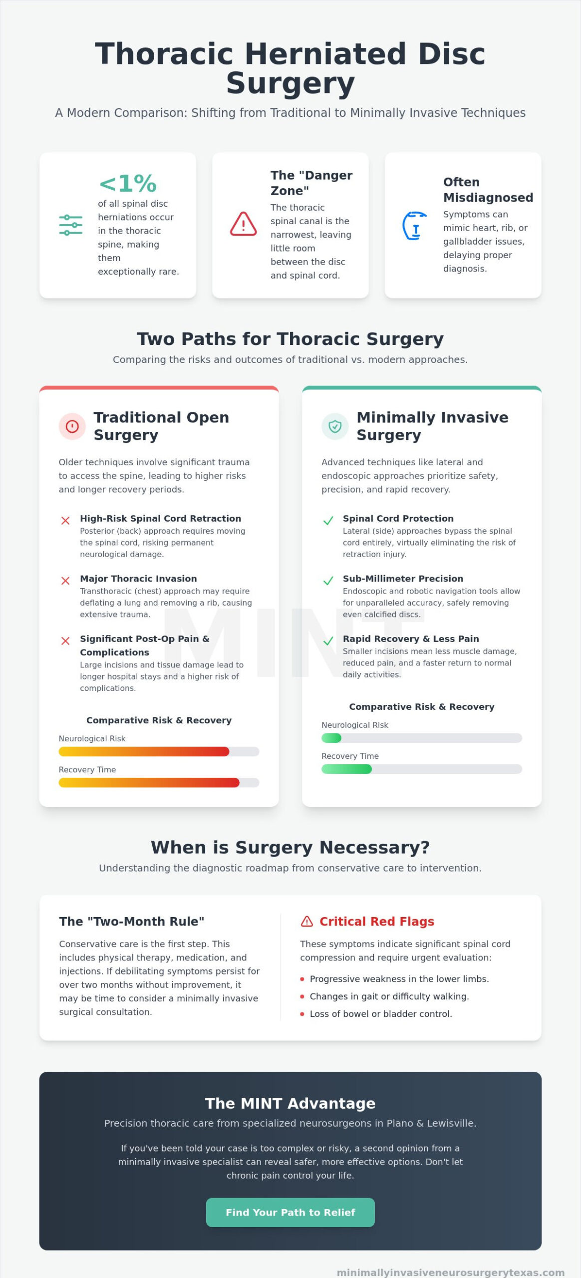
Did you know that thoracic disc herniations account for less than 1% of all spinal disc cases? Because they are so rare, many patients spend months suffering from mid-back pain that mimics heart or rib issues before receiving an accurate diagnosis. It’s completely natural to feel a sense of dread when hearing that thoracic herniated disc surgery is the next step. The proximity of the spinal cord in the mid-back creates high stakes, and traditional open procedures often involve significant physical trauma.
We understand that your primary concern is safety and a return to your normal daily activities. Advanced minimally invasive techniques have shifted the surgical landscape toward precision and rapid recovery. You’ll discover how modern lateral and endoscopic alternatives make thoracic procedures safer and more effective for patients in Plano and Lewisville. This guide provides a clear look at the specific risks of the thoracic region, the latest ultra-minimally invasive options available in 2026, and how to find a qualified neurosurgeon who prioritizes your long-term wellness.
Key Takeaways
- Understand why thoracic herniations are frequently misdiagnosed as heart or rib issues and how to recognize the specific symptoms of a T1-T12 protrusion.
- Compare traditional surgical risks with modern thoracic herniated disc surgery, focusing on how lateral and endoscopic techniques protect the spinal cord.
- Discover the role of advanced robotic navigation in achieving sub-millimeter accuracy for complex mid-back procedures.
- Learn the “two-month rule” for conservative care and the critical red flags that signal when minimally invasive intervention is necessary.
- Find out how specialized neurosurgical expertise in Plano and Lewisville offers a clear path toward relieving chronic pain and returning to normal activity.
What is a Thoracic Herniated Disc and Why is Surgery Unique?
A thoracic herniated disc occurs when the soft inner material of a spinal disc protrudes through the tough outer layer in the T1 through T12 region of your mid-back. Understanding What is a Thoracic Herniated Disc is the first step toward effective treatment, as these injuries are remarkably rare, accounting for less than 1% of all spinal disc cases. Because the rib cage provides a high degree of stability, the thoracic spine doesn’t experience the same wear and tear as the neck or lower back. However, when a herniation does occur here, the stakes for thoracic herniated disc surgery are significantly higher due to the anatomical “danger zone” of the mid-back.
The spinal canal in the thoracic region is significantly narrower than in the cervical or lumbar areas. This means there’s very little buffer space between the disc and the spinal cord. Even a small protrusion can lead to compression, which may cause progressive lower limb weakness or gait changes. Common symptoms include:
- Dull or sharp pain centered in the mid-back.
- Radiating pain that “wraps around” the rib cage.
- Numbness or tingling that travels through the torso.
- Potential dysfunction in bowel or bladder control.
Thoracic vs. Lumbar Herniations
While lumbar herniations typically cause sciatica, which is pain shooting down a single leg, thoracic issues present a more complex diagnostic puzzle. The wrap-around rib pain often mimics internal medical issues like gallbladder disease, heart conditions, or even shingles. Because the thoracic spine has less room for the spinal cord, symptoms can be more bilateral and systemic than the localized pain found in the lower back. This often leads to a long journey for patients in Plano and Lewisville before they find the correct neurosurgical specialist.
Anatomy of the Thoracic Spine (T1-T12)
The twelve vertebrae of the thoracic spine are anchored by the rib cage, which provides essential stability but creates a hurdle for surgical access. The thoracic spinal canal possesses the smallest diameter relative to the spinal cord of any region in the entire vertebral column. Additionally, discs in this area are more prone to calcification over time. This means they can become hardened and more difficult to remove than the softer herniations found elsewhere. This structural complexity is why thoracic herniated disc surgery requires a specialized approach that prioritizes cord protection and spinal stability.
The Challenges of Traditional Thoracic Disc Surgery
Many patients are told by their primary physicians or general surgeons that their condition is “too risky” to operate on. This hesitation stems from the historical challenges associated with traditional thoracic herniated disc surgery. In the past, surgeons primarily utilized the posterior approach, which involves entering through the back. However, because the spinal cord sits directly in the path of the disc, this method requires retracting or moving the cord to reach the herniation. In the narrow thoracic canal, even minimal retraction can lead to permanent neurological deficits. This “Posterior Problem” created a legacy of caution that still influences how many doctors view mid-back surgery today.
To avoid the spinal cord, traditional surgeons often pivoted to the transthoracic approach. This major procedure requires entering through the chest cavity, which frequently necessitates deflating a lung and occasionally removing a portion of a rib to gain access to the spine. While this provides a clear view of the disc, the collateral damage to the patient’s body is extensive. The trauma of opening the chest cavity and the subsequent need for chest tubes often results in significant post-operative pain and a much higher risk of complications compared to procedures in the neck or lower back. If you have been told your case is too complex, consulting a specialist in minimally invasive spine surgery may reveal modern options that bypass these traditional hurdles.
The Risk of Spinal Cord Compression
The blood supply to the thoracic spinal cord is particularly sensitive, often described as a “watershed” zone with less redundant flow than other regions. During traditional surgery, any disruption to this delicate vascular network can have serious consequences. Furthermore, thoracic discs are frequently calcified, meaning they have hardened and potentially adhered to the protective covering of the spinal cord. Safely separating a “stuck” disc from the cord requires extreme precision. Modern surgeons now utilize advanced neurological monitoring to track cord health in real time, ensuring that every movement is within safe parameters.
Traditional Recovery Timelines
Patients undergoing traditional open transthoracic procedures typically face a hospital stay of 3 to 5 days. The recovery process is often hindered by the presence of chest tubes and the lingering soreness from rib resection, making deep breathing and movement difficult in the initial weeks. Full recovery and a return to normal activity can take several months, as the body must heal from both the spinal work and the invasive chest entry. These extended timelines are a primary reason why many patients seek out the more modern, muscle-sparing alternatives available in 2026.
Minimally Invasive Approaches: Lateral and Endoscopic
The evolution of surgical technology has transformed thoracic herniated disc surgery from a high-risk “last resort” into a precise, muscle-sparing procedure. In the past, the trauma of the entry was often as significant as the spinal condition itself. Today, we utilize specialized corridors that allow us to reach the T1-T12 region without the extensive tissue disruption seen in traditional open surgery. By prioritizing the preservation of natural spinal stability, these modern methodologies offer a safer path for patients in Plano and Lewisville to regain their quality of life.
One of the most significant shifts in care is the move toward outpatient or short-stay thoracic procedures. Traditional methods often required multiple days of inpatient monitoring to manage pain and potential complications. With minimally invasive techniques, the reduction in blood loss and internal trauma means many patients are walking within hours of their procedure. This efficiency doesn’t just shorten the hospital stay; it fundamentally changes the trajectory of the patient’s recovery, allowing for an earlier return to normal activity.
The Lateral Access Advantage
The lateral approach represents a major breakthrough in performing thoracic herniated disc surgery. Instead of navigating through the back (posterior) or the chest (transthoracic), the surgeon accesses the disc from the side. By moving between the ribs, we create a direct path to the disc space that avoids the spinal cord entirely. This provides the best possible view of the interface where the disc meets the cord, allowing for a safer decompression. Because the posterior muscles and ligaments remain untouched, the risk of “failed back surgery syndrome” in the thoracic region is significantly lower than with older, more invasive techniques.
Endoscopic Precision
For many patients, endoscopic spine surgery is the gold standard for treating localized herniations. This technique involves a small incision, often less than one centimeter, through which an HD camera and specialized micro-instruments are inserted. The high-definition visualization allows the neurosurgeon to see the compressed nerves with incredible clarity, ensuring that only the problematic disc material is removed. Endoscopic tools are ideal for soft disc herniations because they facilitate targeted removal without the need for extensive bone or muscle resection. This precision minimizes the formation of internal scar tissue, which is a common cause of post-operative stiffness and persistent pain.
Diagnostic Roadmap: When is Surgery Necessary?
Surgery is rarely the first answer for mid-back pain. Most patients find relief through conservative measures, and we typically adhere to the “Two-Month Rule” for non-emergency cases. This protocol involves exhausting options like specialized physical therapy and targeted injections before considering thoracic herniated disc surgery. If your symptoms haven’t improved after eight weeks, or if they begin to worsen despite these treatments, it’s time to evaluate surgical intervention. Surgery is a precise tool designed to restore function when the body’s natural healing process needs assistance.
However, certain “red flags” demand an immediate shift in the diagnostic roadmap. If you experience sudden gait changes, difficulty with balance, or progressive leg weakness, your spinal cord may be under acute stress. Bowel or bladder dysfunction is another critical indicator that the “wait and see” approach is no longer safe. In these high-stakes scenarios, surgical decompression becomes a clinical necessity to prevent permanent neurological damage and ensure a successful return to normal activity.
Recognizing Neurological Decline
Myelopathy is the clinical term for spinal cord dysfunction caused by chronic compression. Unlike localized back pain, which is often muscular or joint-related, myelopathy presents as a loss of coordination or a heavy, “wooden” feeling in the legs. Because the thoracic cord has a sensitive blood supply, early intervention is vital. We aim to intervene before these neurological deficits become permanent, utilizing modern techniques to relieve pressure and preserve the integrity of the spinal cord.
Advanced Imaging in North Texas
Accurate mapping is the foundation of a successful outcome. Standard X-rays are useful for bone alignment, but they often miss the soft tissue pathology of a disc herniation. High-resolution MRI is our primary tool for visualization. For patients with calcified discs, we may also utilize CT myelograms to see the hardened edges of the herniation. This level of detail allows our specialists in minimally invasive neurosurgery to plan a corridor that avoids sensitive structures while maximizing decompression.
When preparing for a consultation in Plano or Lewisville, bring your recent imaging on a disc or ensure it is available via a digital portal. Document the timeline of your symptoms and any treatments you have already attempted. This clarity allows us to build a precise diagnostic roadmap tailored to your specific recovery goals and physical needs. If you’re experiencing mid-back pain that isn’t responding to rest, contact our team today to schedule a comprehensive spinal evaluation.
The MINT Advantage: Precision Thoracic Care in Plano & Lewisville
Dr. Scott Kutz leads Minimally Invasive Neurosurgery of Texas (MINT) with a specialized focus on the rare and complex cases that other practices often decline. While many surgeons find the T1-T12 region intimidating due to its anatomical constraints, Dr. Kutz utilizes a “high-tech healer” approach to make thoracic herniated disc surgery a predictable and successful experience. By operating out of our specialized centers in Plano and our newly relocated main office at 1850 Lakepointe Drive in Lewisville, we provide North Texas patients with access to world-class spinal care without the need to travel to a massive, impersonal hospital system.
We don’t rely on traditional “freehand” techniques for these high-stakes procedures. Our practice integrates the Globus Excelsius robotic navigation system alongside Augmedics Augmented Reality (AR) headsets. This combination allows Dr. Kutz to “see” through the patient’s anatomy in real-time, providing a transparent, 3D view of the thoracic spine without the need for large, traumatic incisions. This level of technological integration ensures that every movement is guided by data and every fragment of disc material is removed with absolute precision.
High-Tech Surgical Navigation
Robotic systems are essential when working within the narrow dimensions of the thoracic spinal canal. These tools provide sub-millimeter accuracy, ensuring that hardware placement and disc removal occur exactly as planned in the pre-operative 3D map. This precision is particularly vital when navigating the “danger zone” near the spinal cord. For patients whose condition requires stabilization, MINT is a recognized leader in minimally invasive spinal fusion Texas, offering a way to secure the mid-back while minimizing blood loss and protecting the delicate vascular supply of the thoracic cord.
Recovery and Results at MINT
The first 48 hours after your procedure are designed for comfort and early mobility. Because we prioritize muscle-sparing corridors and endoscopic techniques, many of our patients are walking within an hour of completing their surgery. We don’t just perform a procedure; we coordinate a personalized recovery protocol that focuses on your specific lifestyle goals. Our team works with you to implement recovery milestones that facilitate a rapid return to normal activity. The MINT promise is built on this blend of compassionate, patient-centered care and a master’s level of surgical precision, giving you the confidence to move forward with your life free from chronic mid-back pain.
Reclaiming Your Quality of Life through Modern Innovation
Modern surgical technology has fundamentally redefined the possibilities for patients suffering from mid-back pain. As we’ve explored, the move toward lateral and endoscopic approaches allows neurosurgeons to bypass traditional “danger zones” while preserving natural spinal stability. By integrating robotic navigation and augmented reality, we achieve a level of precision that ensures thoracic herniated disc surgery is no longer a high-risk last resort, but a safe and effective path to relief.
Board-certified neurosurgeon Dr. Scott Kutz specializes in these advanced robotic and endoscopic spine techniques, providing a sophisticated level of care that prioritizes your safety and long-term wellness. With convenient locations in Plano and Lewisville, our team is dedicated to guiding you through every step of your diagnostic and recovery journey. You deserve a treatment plan that matches the high-tech standards of 2026 while offering the personal attention of a specialized local physician. Request an evaluation for thoracic disc surgery with Dr. Scott Kutz today.
Don’t let the rarity of your condition or the fear of traditional procedures keep you from finding the relief you need. We’re committed to helping you achieve a rapid return to normal activity and a future free from chronic physical limitations.
Frequently Asked Questions
Is thoracic herniated disc surgery considered a major operation?
Traditional open surgery in the thoracic region is considered a major procedure, but modern minimally invasive neurosurgery has significantly reduced the physical impact. By utilizing small incisions and muscle-sparing techniques, we avoid the extensive trauma of opening the chest cavity or deflating a lung. This shift allows for faster healing and a lower risk profile compared to older surgical methods.
What are the success rates for minimally invasive thoracic discectomy?
Success rates for thoracic herniated disc surgery are high, with research indicating success for thoracic discectomies is approximately 80%. Specialized endoscopic procedures have shown success rates as high as 95% in eliminating chronic back pain. These outcomes are more predictable today thanks to the integration of robotic navigation and high-definition visualization.
How long is the recovery period after thoracic spine surgery?
Most patients are walking within an hour after a minimally invasive procedure at our center. While traditional open surgery often required hospital stays of 3 to 5 days, our advanced techniques allow for much shorter stays or even outpatient care. A full return to normal activity typically occurs within a few weeks as the small incisions heal quickly.
Can a thoracic herniated disc cause leg weakness or paralysis?
Yes, because the thoracic spinal canal is the narrowest part of the spine, a herniated disc can directly compress the spinal cord. This pressure can lead to progressive leg weakness, balance issues, or even paralysis if the compression is severe and left untreated. Early intervention is vital to prevent permanent neurological damage when these red flags appear.
Why do some doctors refuse to perform thoracic disc surgery?
Many surgeons hesitate because thoracic herniations are rare, representing less than 1% of all disc cases. The proximity to the spinal cord and the vital organs in the chest makes traditional posterior approaches technically difficult and risky. Our practice specializes in these complex cases by using lateral corridors that reach the disc without disturbing the spinal cord.
What is the difference between a microdiscectomy and a thoracic discectomy?
A microdiscectomy is a common procedure usually performed in the lower back to relieve sciatica. A thoracic discectomy specifically targets the T1-T12 region of the mid-back. Because the anatomy is different, thoracic procedures require specialized surgical corridors, such as lateral or endoscopic entries, to ensure the spinal cord is never retracted or moved.
Will I need a spinal fusion if I have thoracic disc surgery?
A spinal fusion isn’t always necessary for every patient. We prioritize preserving as much natural movement as possible through endoscopic disc removal. However, if the disc herniation has caused instability or requires extensive bone removal, we utilize robotic navigation to perform a precise fusion that stabilizes the mid-back and protects the cord.
Are there non-surgical alternatives for a thoracic herniated disc?
Conservative care is the standard first step for patients who don’t have severe neurological deficits. Non-surgical options include specialized physical therapy, anti-inflammatory medications, and targeted spinal injections. We typically recommend these treatments for at least eight weeks before considering surgical intervention, unless emergency symptoms like bowel or bladder dysfunction are present.