
Endoscopic decompression relieves pressure on spinal nerves through a small incision. Minimally Invasive Neurosurgery of Texas provides Endoscopic Spine Surgery in Lewisville and Plano, TX, under the leadership of Dr. Scott Kutz, a board-certified neurosurgeon. If you’re wondering what endoscopic decompression surgery is, it’s a procedure that uses a small camera and instruments to remove tissue crowding a nerve.
Spinal stenosis, a narrowing of the spinal canal, and a herniated disc can both press on nerves. Pain, numbness, tingling, or weakness may make everyday activities harder, so it’s natural to ask: what is endoscopic decompression surgery, and what might treatment involve?
This guide explains how the procedure works, which conditions it may address, and how its potential benefits compare with other approaches. You’ll also learn what postoperative monitoring and recovery may involve, including a gradual return to daily activities.
Key Takeaways
- Learn how surgeons use endoscopic tools to relieve pressure on spinal nerves.
- See which spinal conditions may be treated and why stability matters when considering decompression.
- Understand how a small incision can limit disruption to surrounding tissue.
- Explore what monitoring, returning to daily activities, and recovery may involve.
- Compare the potential advantages of endoscopic decompression with factors that can affect whether it’s appropriate.
What conditions does endoscopic spine surgery treat?
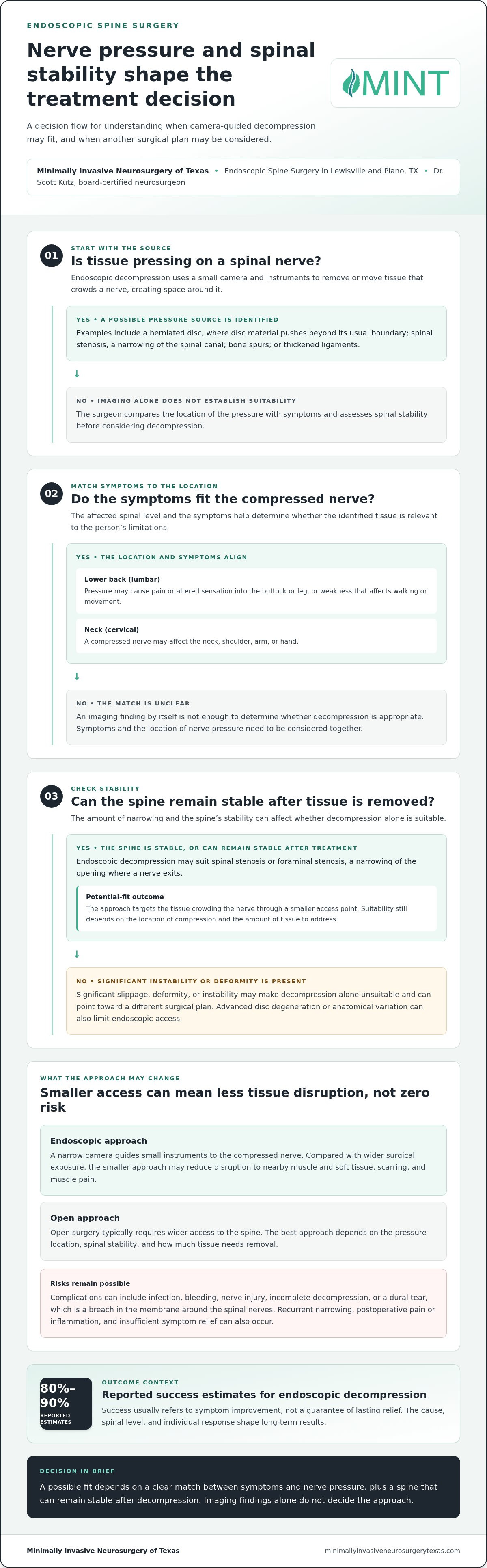
Endoscopic spine surgery targets tissue that presses on spinal nerves, including disc material, bone spurs, or thickened ligaments. Surgeons consider the location of that pressure, the symptoms it causes, and whether the spine can remain stable after decompression.
Stenosis and Herniated Discs
Spinal stenosis means narrowing of the spinal canal, while a herniated disc occurs when disc material pushes beyond its normal boundary. Either condition can crowd nearby nerves and cause pain, numbness, weakness, or difficulty walking. Endoscopic decompression removes or moves the tissue causing pressure, rather than treating an imaging finding alone. The procedure aims to create space around the affected nerve.
Conditions That Compress Spinal Nerves
Bulging discs, facet arthritis, bone spurs, thickened ligaments, scoliosis, and spondylolisthesis can narrow the canal or the openings where nerves exit. Spondylolisthesis means one vertebra slips out of alignment with another. Nerve compression can cause radiating pain, tingling, numbness, or weakness. These symptoms help connect a person’s daily limitations with the spinal level shown on imaging.
Endoscopic decompression most often addresses lumbar problems, meaning conditions in the lower back. Examples include lumbar spinal stenosis and a herniated disc irritating a nerve that travels into the leg. Endoscopic spine surgery can also address cervical conditions in the neck when a disc or narrowed passage compresses nerves. The affected region matters because the surgeon must reach the specific site of pressure while protecting nearby structures.
Symptoms depend on which nerves are under pressure. Lower-back compression can cause pain or altered sensation that travels into the buttock or leg, while weakness can interfere with walking or normal movement. Cervical nerve compression can affect the neck, shoulder, arm, or hand. Bowel or bladder changes can signal a serious neurological problem and require prompt medical attention.
Imaging findings alone don’t establish whether decompression is appropriate. Surgeons compare the compressed nerve’s location with the patient’s symptoms, then assess spinal stability and the extent of narrowing. Severe instability or deformity can make decompression alone unsuitable, because removing tissue could leave the spine unable to remain stable. Advanced degenerative disc disease or anatomical variations can also limit endoscopic access.
Endoscopic decompression generally suits spinal stenosis or foraminal stenosis, which narrows the opening where a nerve exits, when the spine remains stable or can remain stable after treatment. Significant slippage, deformity, or instability can point toward a different surgical plan. The endoscopic spine surgery approach focuses on relieving nerve pressure through a smaller access point.
Potential benefits of micro-invasive decompression
Endoscopic decompression uses a camera and small instruments to relieve pressure on a nerve through limited access. Its potential benefits come from reducing disruption to surrounding tissue, not from eliminating surgical risks.
Benefits of Minimally Invasive Surgery
Minimally invasive surgery reaches a treatment area through smaller openings and aims to limit damage to nearby muscles and soft tissue. In endoscopic spine surgery, the surgeon views the area through a camera and removes tissue compressing a nerve. Less tissue disruption may mean less muscle pain and minimal scarring. These potential differences may matter to people weighing surgery against ongoing symptoms.
Benefits Compared With Open Surgery
Open surgery typically requires wider access to the spine, while endoscopic decompression uses a small incision and specialized instruments. The smaller approach can reduce tissue disruption, scarring, and muscle pain compared with wider surgical exposure. It doesn’t make every case suitable for endoscopy, however. The location of compression, spinal stability, and the amount of tissue to remove all affect the approach.
During the procedure, the surgeon guides an endoscope, a narrow camera, toward the compressed nerve and views the treatment area. Small instruments remove or move the tissue causing pressure, while irrigation keeps the view clear. Anesthesia may involve general anesthesia or local anesthesia with sedation, depending on the case. The goal is targeted decompression, or creating space around the nerve.
Potential benefits don’t eliminate risks. Complications can include infection, bleeding, nerve injury, a dural tear, or incomplete decompression. A dural tear is a breach in the membrane surrounding the spinal nerves. Recurrent stenosis, postoperative pain or inflammation, and insufficient symptom relief can also occur. Patients can discuss how the location and extent of their nerve compression affect these risks.
Reported success estimates for endoscopic decompression range from 80% to 90%. Success usually refers to improvement in symptoms, rather than a guarantee of lasting relief. The underlying cause, the affected spinal level, and the patient’s response shape long-term results. A clear discussion of the expected outcome should distinguish pain relief from changes in numbness, weakness, or daily function.
Before surgery, patients can ask which tissue is compressing the nerve, what the procedure will remove, and why endoscopic access fits their anatomy. They can also ask about anesthesia, possible complications, and which symptoms the operation aims to improve. Those questions help clarify the plan and its limits. The endoscopic spine surgery approach uses camera-guided access to treat selected sources of nerve pressure.
Small-incision and endoscope dimensions
Endoscopic access combines a skin incision, a working channel, a camera, and surgical instruments. The exact dimensions vary by technique, so incision length and tube diameter describe different parts of the approach.
Some endoscopic lumbar decompression approaches use a tube measuring 8 mm in diameter; others describe a 10.5 mm tube. A micro-laminotomy, a small opening in the bone at the back of the spine, may use a 22 mm tube. These examples represent distinct access methods, not a single standard size for every procedure.
One described endoscopic decompression incision measures less than one-quarter inch. Another approach may use an incision smaller than 1 cm. The skin opening provides the entry point, while the tube creates a working passage through soft tissue toward the treatment site. A larger tube does not necessarily mean a similarly sized skin incision, because instruments pass through the access channel.
Before placing instruments, the surgeon identifies the entry point with imaging guidance. Fluoroscopy, which provides live X-ray images, or intraoperative CT, which creates cross-sectional images during surgery, can help mark the route. X-ray guidance also helps track instruments as they approach spinal structures. Real-time radiographic images provide another view of their position.
After marking the route, the surgeon uses muscle dilators to gently separate tissue and create space for the tube. This approach separates muscle fibers rather than cutting through muscle. The endoscope carries a camera view to a monitor, while irrigation keeps the surgical field visible. Small instruments work through the channel to address tissue compressing nerves, such as herniated disc material or bone.
The camera’s close view helps the surgeon see the target through a limited opening. In lumbar surgery, access may come through the interlaminar space, an opening between bones at the back of the spine. Biportal endoscopic surgery uses one incision for the endoscope and another for instruments. This differs from a uniportal approach, where the camera and tools share one access route.
Incision size alone doesn’t describe the full surgical approach. A laminectomy, which removes part of the lamina or back portion of a vertebra, may use an incision measuring 1 to 2 inches, depending on treated levels. A spinal fusion incision may measure 2 to 3 inches or more. Endoscopic methods use narrower access, but the surgeon still plans around nerves, bone, and the spinal cord.
These measurements help explain how instruments reach the spine, not which technique suits every case. The target level, tissue to remove, and route to that tissue shape the chosen access. For example, a surgeon may select a different approach for a disc herniation than for bone narrowing around a nerve.
Recovery timelines and hospital stays
After endoscopic decompression, many patients return to light daily activities within a few days, while full activity usually takes around six weeks. Recovery starts with short walks and gradual movement. The treated spinal level and procedure shape each stage.
For spinal stenosis, clinicians monitor patients for a few hours after surgery. Endoscopic spine surgery usually takes place on an outpatient basis, so patients typically leave the hospital the same day. Monitoring allows the care team to observe the immediate postoperative period before discharge. Its duration depends on how the patient progresses after the procedure.
Walking shortly after surgery helps maintain movement during recovery. Patients can resume light activity the next day, then gradually increase activity over seven days after endoscopic lumbar decompression. This gradual pace supports a return to daily routines without jumping straight into strenuous tasks. Desk-work schedules differ from jobs that involve lifting, bending, or prolonged physical effort.
Many patients return to desk work within one week. They should avoid heavy lifting and strenuous activity for a few weeks after endoscopic decompression. Surgeons may also recommend limiting bending or twisting, and some patients wear a back brace for several weeks. These restrictions protect the surgical area as patients rebuild normal movement.
Physical therapy can support recovery by helping patients regain movement and build tolerance for activity. Timing and exercises should match the procedure and the patient’s progress. Patients can ask their care team when to begin therapy, which movements to limit, and how to increase walking or other activity. For individuals managing gait adjustments or lower-limb discomfort as they get back on their feet, allied health providers like Active Foot Care show how targeted foot and mobility support can assist overall recovery. Those details turn general recovery milestones into a practical plan.
Traditional procedures can involve different timelines. After a laminectomy, patients may spend one day in the hospital, and limited activities may resume after four to six weeks. Spinal fusion often involves a one- to two-day hospital stay, with full recovery taking four to six months. Fusion joins spinal bones, so recovery differs from decompression without fusion.
After discharge, follow the surgical team’s instructions for activity, wound care, and follow-up visits. Report new or worsening symptoms promptly, especially changes that interfere with movement or daily tasks. Keep a simple record of walking, work, and pain levels. These observations help the care team assess progress and adjust activity guidance during recovery.
Advantages of endoscopic decompression
Endoscopic decompression uses a camera and small instruments to target tissue pressing on spinal nerves. Its central advantage is focused access: the surgeon treats the source of compression without placing spinal hardware. For a patient with a stable spine and a localized herniated disc or stenosis, that distinction can shape the surgical plan.
Smaller access can reduce disruption to surrounding tissue compared with wider surgical exposure. Endoscopic procedures can cause less tissue damage, minimal scarring, and reduced muscle pain. These potential benefits matter because muscle soreness and incision discomfort can affect early movement, including walking and everyday activities. They describe possible advantages of the approach, not a promise of symptom relief.
The camera gives the surgeon a close view of the treatment area through a narrow access route. Small instruments can remove disc material, bone, or other tissue compressing a nerve. This focused view supports targeted decompression: creating space around the affected nerve. The anatomy and location of pressure guide what the surgeon can safely address.
Endoscopic decompression also differs from procedures that place hardware. It doesn’t involve hardware placement and aims to relieve nerve pressure by removing the compressive tissue. That may be relevant when the spine remains stable and decompression alone can address the problem. Significant instability or deformity can change the surgical decision because the spine may need more than pressure relief.
Recovery can reflect the smaller access and reduced muscle disruption. After endoscopic decompression for spinal stenosis, patients usually spend a few hours under observation, and many resume light daily activities within a few days. Full activity usually returns around six weeks after endoscopic spine surgery. These milestones can help with planning, while activity increases gradually during healing.
Weigh potential benefits against the goal of treatment. Decompression targets nerve-related symptoms such as radiating pain, numbness, or weakness; it doesn’t correct every source of back pain. A candidate needs a clear match between symptoms, imaging, and the tissue causing compression. For example, removing a herniated disc fragment may relieve pressure, while advanced instability can make decompression alone a poor fit.
The practical takeaway is to compare the method with the problem it must solve. A small incision and camera-guided access may limit tissue disruption, but the surgeon still needs to reach the correct level and protect spinal stability. Ask how the proposed procedure addresses the specific nerve compression, which symptoms it targets, and what activity limits follow. This discussion connects potential advantages with your treatment goals.
Minimally Invasive Neurosurgery of Texas: Minimally Invasive Spine Surgery
Minimally Invasive Neurosurgery of Texas provides minimally invasive spine care for patients exploring treatment for spinal nerve compression. Dr. Scott Kutz, a board-certified neurosurgeon and Fellow of the AANS and ACS, emphasizes matching treatment discussions to a clear diagnosis and the patient’s symptoms.
If back or leg symptoms persist, a discussion can clarify whether Endoscopic Spine Surgery fits the affected spinal level and the source of pressure. Patients can also discuss other treatment options, including Artificial Disc Replacement when appropriate. Contact the practice to discuss your spine care.
Prepare for a Clear, Informed Next Step
Understanding what endoscopic decompression surgery involves can help you have a more useful conversation about your care. Before the discussion, note which activities your symptoms disrupt, what movements bring relief or discomfort, and what you hope to regain. Specific examples, such as difficulty sitting through work or walking comfortably, can help focus the conversation on your priorities.
Use the visit to clarify how the proposed plan fits those priorities. Ask what changes the procedure aims to address, how the care team will assess progress, and what other treatment paths may suit your situation. Clear answers can help you weigh the benefits and limits of surgery while keeping your daily goals in view.
If you’re ready to discuss your spine concerns and possible next steps, contact Minimally Invasive Neurosurgery of Texas. A thoughtful conversation can help you move forward with greater clarity and confidence.
Frequently Asked Questions
What is an Endoscopic Decompression?
Endoscopic decompression is a surgical treatment focused on relieving pressure around a spinal nerve. The practical distinction is that the surgeon uses a camera-guided approach rather than viewing the treatment area through a wider opening. Before deciding, clarify which imaging finding matches your symptoms and what change the operation aims to achieve.
How long will the procedure take?
The procedure typically takes 2-3 hours, depending on its complexity. Ask whether the estimate includes only the operation or also preparation and post-surgical monitoring. The number of spinal levels and the work required at each level can affect the plan. Knowing what the time estimate covers can help you arrange transportation and support for the day.
What is the recovery like?
Recovery involves following activity guidance and noticing how symptoms change as you resume routines. Track which daily tasks feel easier or remain difficult, such as sitting at a desk or walking. Share those observations during follow-up. They can help the care team understand your progress and guide decisions about increasing activity or adjusting restrictions.
Who is a good candidate for endoscopic spinal surgery?
A strong potential fit has a defined surgical target and a reason the chosen access method can reach it. During evaluation, discuss how the proposed treatment relates to your specific symptoms, imaging, and spinal anatomy. Bring a concise history of prior spine treatments and the activities your symptoms limit. That context helps make the discussion about candidacy specific to your situation.
What is endoscopic decompression surgery and how does it work?
The surgeon uses an endoscope, a narrow camera, to view the treatment area while working through a small access route. Surgical instruments address the identified source of nerve pressure. Ask which part of the procedure requires the most precision in your case, how the surgeon will confirm the target, and what change the operation aims to make to your symptoms.
What conditions is endoscopic decompression surgery used to treat?
Surgeons may use the procedure when spinal narrowing or disc material puts pressure on a nerve. Location matters: lower-back nerve compression can affect the leg, while compression in the neck can affect the arm or hand. Ask the surgeon to identify the affected level and explain how it relates to your pattern of symptoms.
Who is a good candidate for endoscopic decompression surgery?
Candidate selection depends on more than a diagnosis label. A useful consultation connects the planned treatment to the patient’s symptoms, imaging, anatomy, and goals. Ask what findings support the proposed approach and what might change the plan. This can clarify why the surgeon recommends decompression, another treatment, or continued nonsurgical care.
How does endoscopic decompression compare with traditional open surgery?
The approaches differ in how the surgeon accesses the spinal treatment area. Endoscopic surgery uses a camera and narrower access, while open surgery uses wider exposure. The more useful comparison is how each method addresses the specific surgical target and surrounding anatomy. Ask which differences matter most in your case, including the planned treatment and expected activity restrictions.
This information is for educational purposes only and isn’t a substitute for professional medical advice, diagnosis, or treatment. The website and its content are provided “as is.”