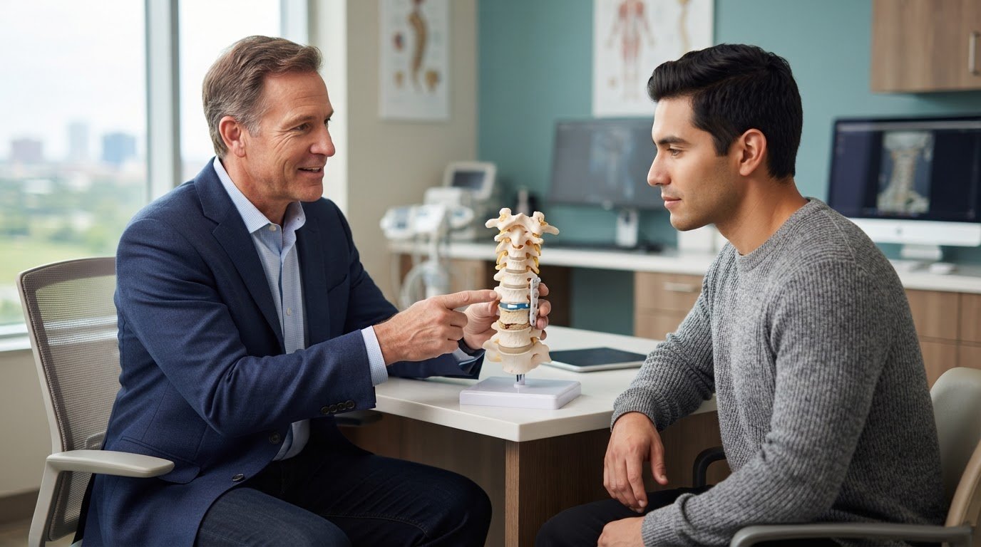
Minimally Invasive Neurosurgery of Texas provides minimally invasive spine surgery in Lewisville and Plano, Texas. Board-certified neurosurgeon Dr. Scott Kutz leads the practice’s spine care.
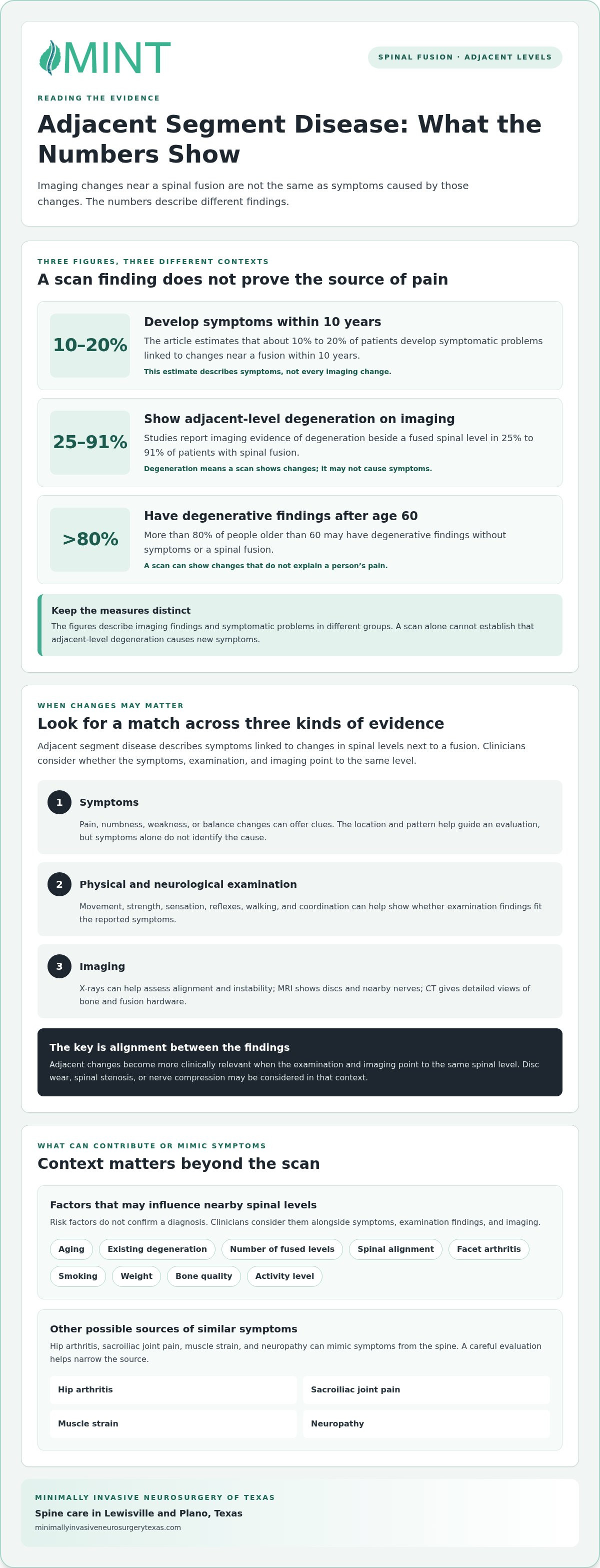
About 10% to 20% of patients develop symptoms linked to changes near a spinal fusion within 10 years. Doctors use adjacent segment disease to describe those symptoms, while imaging can show degeneration without pain.
New pain after fusion can be concerning, but several conditions can cause similar symptoms. This guide explains adjacent segment disease, its possible symptoms and causes, and conditions that can mimic it, so you know what an evaluation may consider.
Key Takeaways
- Imaging changes beside a fusion don’t always cause symptoms; the distinction matters when interpreting scan results.
- New pain, numbness, weakness, or balance changes can offer clues, but they don’t identify the cause on their own.
- Fusion-related changes in spinal mechanics may contribute to stress on nearby levels, alongside factors such as existing degeneration and spinal alignment.
- Hip arthritis, sacroiliac joint pain, muscle strain, and neuropathy can mimic symptoms from the spine, so a careful evaluation helps narrow the source.
- Understanding how symptoms, examination findings, and imaging fit together can help clarify which treatment options may be appropriate.
What Is Adjacent Segment Disease?
Adjacent segment disease describes symptoms linked to changes in spinal levels next to a fusion. The distinction between scan findings and symptoms helps explain what those changes mean for you.
Nonsurgical care may include exercise, physical therapy, and epidural steroid injections, which deliver anti-inflammatory medicine near spinal nerves. For those exploring non-invasive management options, integrative practices such as Dr. Philip Boamah | Natural Medicine & Physical Therapy highlight how targeted physical therapy can help patients address joint and muscle pain. These approaches may suit mild to moderate symptoms when there is no major weakness or instability.
Degeneration Versus Symptomatic Disease
Adjacent segment degeneration means imaging shows changes beside a fused spinal level; disease means those changes cause symptoms. This difference matters because an X-ray, CT scan, or MRI can reveal degeneration without identifying the source of pain.
Imaging studies report adjacent-level degeneration in 25% to 91% of patients with spinal fusion. Degenerative findings also appear in more than 80% of people older than 60 without symptoms or fusion. A scan alone therefore cannot establish that degeneration explains new pain.
Changes become clinically relevant when examination findings and imaging point to the same spinal level. They may include disc wear, spinal stenosis, or nerve compression. Stenosis is narrowing around the spinal cord or nerves; compression is pressure on those structures. Matching these findings with symptoms helps distinguish symptomatic disease from an incidental scan result.
Risk Factors for Adjacent Segment Disease
Aging and degeneration that existed before fusion can contribute to changes at nearby levels. Spinal wear may progress, and existing disc or joint problems can continue after surgery. New symptoms may therefore reflect both ongoing degeneration and altered mechanics, rather than a single cause.
Risk can also vary with the number of fused levels, spinal alignment, facet arthritis, smoking, weight, bone quality, and activity level. Facet arthritis affects the small joints that guide spinal movement. These factors may influence how neighboring segments handle movement and load.
Risk factors alone don’t confirm a diagnosis. For example, someone with pre-existing disc wear may show changes on an MRI without developing new pain. Another person may have radiating pain or numbness if a nearby level irritates a nerve. Clinicians consider risk alongside symptoms and examination findings.
Adjacent Segment Changes After Fusion
Spinal fusion joins vertebrae so they no longer move independently at the treated level. The segments above or below may take on additional work to support movement. Over time, this shift can contribute to degeneration, particularly when nearby levels already show wear.
Changes can appear months or years after surgery. About 10% to 20% of patients develop symptomatic problems within 10 years of fusion. This estimate describes symptoms, not every imaging change, and adjacent-level problems don’t automatically mean the original fusion failed.
When symptoms arise, clinicians compare a patient’s history and examination with imaging of the fused and neighboring levels. X-rays, MRI, or CT scans may help assess alignment, narrowing, and pressure on nerves. Comparing these findings helps distinguish a symptomatic adjacent level from ordinary degeneration or another source of pain.
Definition and symptoms of adjacent segment disease
Symptoms depend on which nearby spinal level changes and whether the change affects a nerve or the spinal cord. Neck and lower-back problems can therefore cause different patterns of pain, weakness, and difficulty moving.
Cervical and Lumbar Symptoms
Cervical symptoms affect the neck; lumbar symptoms affect the lower back. In the neck, nerve irritation can cause pain that travels to the shoulder, arm, or hand, as well as tingling, numbness, or weaker grip. Radiculopathy describes symptoms caused by an irritated or compressed nerve root.
Pressure on the spinal cord can cause myelopathy, or impaired spinal cord function. Clues may include worsening balance, reduced hand coordination, or difficulty with fine motor tasks. Lumbar symptoms may include low-back pain, buttock or leg pain, numbness, weakness, and reduced walking tolerance.
The location and pattern of symptoms help guide an evaluation. Pain traveling from the lower back into one leg, for example, differs from isolated back soreness; hand clumsiness differs from neck pain alone. New or worsening weakness, balance problems, or bowel or bladder changes need prompt medical assessment.
Physical Examination Findings
A physical and neurological examination checks whether symptoms match changes in movement, strength, sensation, or reflexes. A clinician may assess walking, hand coordination, grip, and limb strength, then compare those findings with the patient’s history and prior surgery records.
Each finding provides context, but none establishes a diagnosis by itself. Leg weakness paired with radiating lumbar pain, for example, may direct attention to a nerve, while balance changes and reduced hand control may raise concern about spinal cord involvement. The examination helps identify which level needs closer assessment.
Describe where symptoms travel, which activities worsen them, and whether they affect walking or hand use. These details can help distinguish a consistent nerve pattern from pain that stays in one area. A clear timeline also matters: new symptoms after a period of improvement differ from discomfort that has remained unchanged since surgery.
Imaging and Symptoms
X-rays, MRI, and CT scans show different features of the spine. Standing or movement-based X-rays can help assess alignment and instability; MRI shows discs and nearby nerves; CT provides detailed views of bone and fusion hardware.
Imaging matters most when the findings match symptoms and examination results. A scan may show narrowing or disc changes without explaining a patient’s pain. In contrast, a nerve compressed at a specific level may fit symptoms that travel into an arm or leg. Diagnosis depends on how the evidence fits together, not on the image alone.
Bring prior imaging and surgery records to an evaluation when available. Comparing older and newer scans can show how a spinal level has changed, while current images help assess nerve pressure, bone anatomy, and fusion status. This comparison supports a clearer explanation of symptoms and next steps.
Why Can It Happen After Spinal Fusion?
Fusion changes movement at the treated level, so neighboring spinal joints may carry more workload. Surgical planning and individual factors can also influence degeneration above or below the fused area.
Patient-Related Risk Factors
Aging and pre-existing disc wear can contribute to later changes near a fusion. Spinal degeneration may continue over time, even when the original surgery treated a different level. For example, a disc that already showed wear before surgery may remain vulnerable to further change.
Risk factors also include smoking, excess weight, bone quality, diabetes, facet arthritis, and activity level. Smoking may be one factor associated with cervical adjacent segment disease. Cervical cases also occur more often in patients younger than 60, while lumbar and cervical areas face greater risk than the thoracic spine.
Weight can increase the load on nearby levels, while weak bone may affect healing and surgical risk. Diabetes, nicotine use, medications, prior infections, and previous surgeries can also shape recovery. These factors help explain why people who undergo similar fusion procedures may have different outcomes.
Surgical Planning Considerations
Surgical planning considers fusion length, spinal alignment, fusion type, and protection of nearby facet joints. These joints guide spinal movement; disrupting them during surgery can add stress to an adjacent level. Alignment matters, too, because an imbalance may shift forces across the spine.
For cervical fusion, placing an anterior plate close to the neighboring disc can increase the risk of adjacent-level ossification, meaning bone formation near that disc. A plate-to-disc distance greater than 5 mm reduces that ossification risk, but does not change the risk of adjacent-segment degeneration itself.
Fusion length presents another planning consideration. Fusing fewer than 3 segments is listed as a risk factor for cervical adjacent-level disease. The number of levels treated must still match the spinal problem, and planning also weighs existing degeneration, stability, and the patient’s anatomy.
Minimally invasive and endoscopic procedures can spare adjoining muscles and ligaments. Limiting disruption to surrounding tissue may reduce one contributor to degeneration, but these approaches don’t eliminate the effects of aging, existing disc wear, or spinal mechanics.
Standing X-rays and movement-based images can help assess alignment and instability before or after spine surgery. Surgeons also consider bone density, nicotine use, weight, and prior procedures when evaluating surgical risk. Patients who smoke or vape should stop before surgery and remain nonsmokers afterward, because nicotine use can affect healing.
Some contributors can change, while others, such as age and existing degeneration, cannot. Addressing nicotine use, weight, and spinal alignment may help reduce risk, but no single planning decision explains every later change. A patient’s anatomy, fusion details, and healing all contribute.
Q: What advice do you give your patients after they undergo a fusion?
After spinal fusion, keep track of new pain, numbness, weakness, limits on walking, or changes in hand coordination. These symptoms don’t prove a complication, but their location and effect on daily activities can help guide an evaluation.
Surgery may be considered when symptoms, examination findings, and imaging identify the same affected level and symptoms impair function. For lumbar disease, surgeons may consider surgery when nonsurgical treatment hasn’t helped or cauda equina syndrome occurs, a serious condition involving pressure on nerves that control the legs and bladder. Severe myelopathy, or spinal cord dysfunction, also requires surgical treatment.
Risk estimates vary by spinal region: the reported annual risk is 2% to 14% after lumbar fusion and 3% after cervical fusion. Aging and fusion may both contribute to changes, although whether fusion accelerates degeneration remains controversial. These figures describe risk across groups, not an individual’s expected outcome.
The operation depends on the problem at the adjacent level. Decompression removes tissue pressing on nerves and may relieve pressure without extending a fusion in selected cases. Surgeons avoid decompression alone when instability already exists or the procedure would create it. Extending a fusion may address instability, deformity, severe disc collapse, or the need for broader nerve decompression.
For selected cervical cases, artificial disc replacement preserves movement at the treated level. Clinicians consider this option based on the affected level, spinal stability, and source of symptoms. Motion-preserving surgery differs from extending a fusion, which joins another level and changes its movement. The choice depends on the anatomy and the specific problem surgery needs to address.
Recovery varies with the procedure, the number of treated levels, and overall health. Follow the surgical team’s activity guidance, and increase walking or other movement according to those instructions. Don’t use someone else’s recovery schedule as your own: a procedure involving one level may call for a different plan than a longer revision operation.
Ask which symptoms need prompt attention and which activities fit your recovery plan. New weakness, worsening balance, loss of hand coordination, bowel or bladder changes, or groin numbness need prompt medical assessment. Track changes in pain, walking distance, and daily tasks. These details help clinicians judge whether symptoms are improving or limiting function.
Outcomes depend on whether the chosen procedure addresses the source of pain and relieves nerve pressure without creating instability. Recovery timelines vary with surgical complexity, treated levels, and health. Surgery aims to improve symptoms and function, but risks and expected recovery depend on the specific spinal problem and treatment plan.
Conditions that can mimic adjacent segment disease
New pain after fusion doesn’t always come from the adjacent spinal level. Hip arthritis, sacroiliac joint pain, muscle strain, neuropathy, and other spine conditions can cause overlapping symptoms. Distinguishing among these causes matters because revision surgery treats a specific spinal problem, not pain alone.
Other Sources of Pain After Fusion
Hip arthritis can cause pain around the hip, while sacroiliac joint pain comes from the joint connecting the pelvis and spine. Muscle strain can cause localized soreness, and neuropathy, or nerve damage, can cause pain, tingling, or numbness. These conditions may occur after an older fusion without indicating a problem at the adjacent level.
Other spinal conditions can also resemble adjacent-level disease. A new herniated disc, for example, may irritate a nerve and cause pain that travels into an arm or leg. Spinal stenosis, or narrowing around spinal nerves, can also produce symptoms similar to those linked with degeneration near a fusion.
How Clinicians Separate the Causes
A useful assessment connects the location and timing of symptoms with examination findings and imaging. Clinicians review the fusion history, examine strength and sensation, and consider whether symptoms follow a nerve pattern. They may also assess hip and pelvic movement when those areas could contribute to pain.
Imaging is most useful when it matches the clinical picture. X-rays can show alignment and movement; MRI can reveal discs and nerve compression; CT can clarify bone anatomy and hardware. A finding at an adjacent level may be incidental if symptoms and examination findings point elsewhere.
For example, a scan may show degeneration next to a fusion while the examination suggests another source of pain. The scan alone doesn’t establish the cause. Before recommending treatment, clinicians compare the affected level with the patient’s pain pattern, neurological findings, and daily limitations.
Why the Distinction Matters for Treatment
Conservative treatment may address symptoms without another procedure when surgery isn’t needed. The right approach depends on the source of pain. Treating nerve compression at an adjacent level won’t resolve pain caused by hip arthritis or muscle strain.
Revision surgery means another operation on the spine. Before considering it, clinicians look for agreement among symptoms, examination findings, and imaging. Decompression may relieve nerve pressure in selected cases, while extending a fusion may address instability or deformity. Each procedure addresses a different structural problem.
Describe where your pain travels, which movements bring it on, and whether it comes with numbness or weakness. Bring prior surgery records and imaging to the evaluation. These details can help distinguish spinal degeneration from a condition that mimics it and clarify whether the next step should address the spine or another source.
Minimally Invasive Neurosurgery of Texas: Minimally Invasive Spine Surgery
Minimally Invasive Neurosurgery of Texas evaluates spine conditions that may cause new pain or nerve symptoms after fusion. Dr. Scott Kutz, a board-certified neurosurgeon and Fellow of the AANS and ACS, serves patients in Lewisville and Plano, Texas.
An evaluation considers your symptoms, examination findings, prior surgery, and imaging before discussing treatment. When a spinal condition calls for surgery, minimally invasive spine techniques may offer an option; complex cases may need a different approach. The appropriate procedure depends on the affected level, spinal stability, and the source of symptoms.
If new or worsening symptoms affect daily activities, contact Minimally Invasive Neurosurgery of Texas to discuss an evaluation.
Take the Next Step Toward a Clearer Plan
New or changing symptoms after fusion can raise difficult questions. As a next step, note when they began, which activities affect them, and how they change your daily routine. This context can help guide the right clinical evaluation, rather than relying on an imaging finding alone.
If you’re concerned about adjacent segment disease, bring your questions and available spine records to an evaluation with a neurosurgical team. Minimally Invasive Neurosurgery of Texas assesses spine-related concerns and discusses whether minimally invasive spine surgery or another treatment approach fits the findings. Dr. Scott Kutz and his team can explain the options clearly, including when surgery may not be appropriate.
Contact Minimally Invasive Neurosurgery of Texas to discuss your concerns and possible next steps. A clearer understanding can help you move forward with greater confidence.
Frequently Asked Questions
How do changes develop over time?
Most symptomatic cases emerge within 5-10 years after the initial fusion, although symptoms can begin sooner or later. This timing can help frame follow-up, but it can’t identify the cause by itself. For example, a new symptom years after surgery still needs assessment alongside your health history and current findings. The timing offers context, not proof that the fused level caused the problem.
Can artificial disc replacement lower risk compared with fusion?
Artificial disc replacement preserves movement at the treated cervical level and may place less stress on nearby levels than fusion. However, that potential advantage doesn’t make it a universal substitute or guarantee that later problems will be prevented. The procedure treats a specific disc condition. Whether motion preservation is suitable also depends on the affected level and your spinal anatomy.
How can patients lower their risk?
Before surgery, discuss bone density, diabetes, medications, prior infections, and previous operations with your care team. These factors can affect surgical risk, healing, and recovery. Patients who smoke or vape should stop before surgery and remain nonsmokers afterward. After surgery, follow your procedure’s activity limits and follow-up plan, and report meaningful changes rather than abruptly changing your activity.
How common is repeat surgery after fusion?
One study reported a 2.6% rate when it counted repeat surgery for adjacent-level problems. That figure measures reoperation, not everyone who develops symptoms or imaging changes. These are different outcomes: a person may have symptoms without needing another operation. Ask which outcome a statistic measures, and whether it applies to your spinal region and type of fusion.
What causes spinal joints to wear?
Spinal joints can wear as their cartilage and surrounding structures change over time. Facet arthritis affects the small joints that guide vertebral movement. Bone formation near a neighboring disc, called adjacent-level ossification, can occur alongside degeneration, but it is a distinct finding. This matters because bone formation and joint wear don’t necessarily explain symptoms in the same way.
How does imaging-based degeneration differ from symptomatic disease?
Imaging-based degeneration describes visible structural changes; symptomatic disease means clinical symptoms are linked to those changes. In the cervical spine, clinicians look for radiculopathy, or nerve-root symptoms, or myelopathy, or spinal cord dysfunction, that matches the motion segment next to the fusion. This clinical context helps distinguish a relevant finding from an unrelated scan observation.
What factors contribute to symptoms at a nearby spinal level?
Researchers link adjacent-level changes to both the progression of existing degeneration and altered spinal mechanics after fusion. However, whether fusion itself accelerates natural progression remains controversial. A change that appears after surgery doesn’t, on its own, show that surgery caused it. Clinicians consider the patient’s prior condition alongside current findings.
Which signs may help identify the affected spinal region?
In the cervical spine, the lower three segments, C4-5, C5-6, and C6-7, most commonly show adjacent-level disease. The affected level can help clinicians interpret where symptoms arise, but a segment name alone doesn’t identify the cause. Share the location and progression of symptoms, and bring prior imaging when available, so clinicians can compare the reported changes with the relevant spinal level.