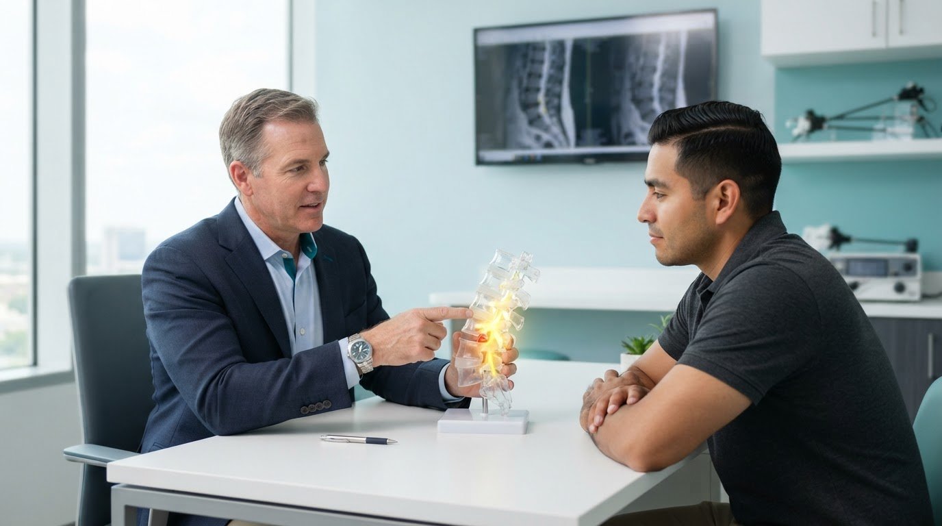
Minimally Invasive Neurosurgery of Texas provides minimally invasive spine surgery in Lewisville and Plano, TX. Board-certified neurosurgeon Dr. Scott Kutz treats spinal conditions, including herniated discs. For people considering minimally invasive lumbar discectomy in Lewisville, TX, the procedure removes or trims disc material pressing on a nerve.
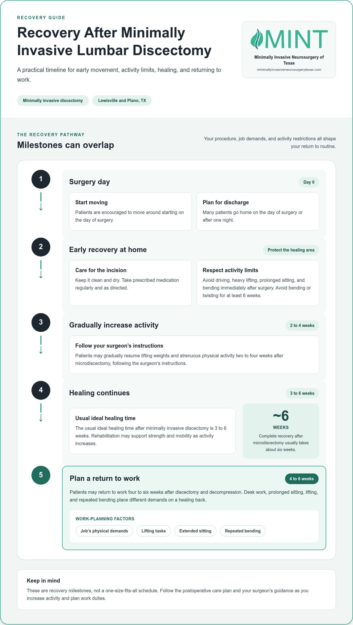
It’s understandable to want to know when you can return to work and daily routines. The usual ideal healing time after minimally invasive discectomy is 3 to 6 weeks, and patients may resume work four to six weeks after discectomy and decompression. Your job’s physical demands can shape return-to-work planning.
This guide explains how surgeons use microdiscectomy to remove disc material pressing on a nerve, how minimally invasive techniques work, and what to expect during recovery, including activity restrictions and a gradual return to routine.
Key Takeaways
- Learn when a herniated disc and persistent nerve symptoms may make surgery a consideration after nonsurgical care.
- Recognize how pain, tingling, numbness, or weakness can point to irritation of nerves in the lower back.
- See how a microdiscectomy uses a focused approach to remove disc material pressing on a nerve.
- Understand why recovery plans account for movement, lifting, bending, and the physical demands of your job.
- Explore how imaging findings and symptoms help guide decisions about treatment and return to normal activity.
What is Minimally Invasive Lumbar Discectomy?
Minimally invasive lumbar discectomy removes or trims herniated disc material that presses on a spinal nerve. The surgeon uses a smaller incision and specialized instruments to reach the affected area.
Benefits of Minimally Invasive Surgery
Minimally invasive spine surgery uses a focused route to reach the problem area while limiting disruption to nearby muscle and other tissue. Compared with an open approach, it can involve less postoperative pain, bleeding, tissue damage, and recovery time. These differences matter when considering how the operative approach may affect comfort and everyday activities after surgery.
Minimally Invasive and Open Discectomy
Both approaches remove disc material compressing a nerve, but differ in how the surgeon reaches the spine. Minimally invasive discectomy uses a smaller incision and specialized instruments; open discectomy requires a wider surgical opening. The minimally invasive approach can reduce tissue disruption and postoperative pain, but the right method depends on the location and cause of nerve compression.
Herniated discs are a common reason surgeons consider lumbar discectomy. A herniated disc occurs when disc material pushes beyond its usual boundary and irritates or compresses a nearby nerve. Spinal stenosis, a narrowing of the spinal canal, can also put pressure on nerves. In that case, a surgeon may perform decompression to create more space around them. Imaging findings need to match a patient’s symptoms to help identify the source of pain.
Treatment typically begins with nonsurgical options to relieve symptoms. These may include physical therapy, exercise, medication, weight loss, or injections. For example, someone whose leg pain interferes with daily activities may try a guided exercise program and medication before considering surgery. Surgery may enter the discussion when conservative care does not provide adequate relief and the clinician can identify the spinal structure causing the symptoms.
To assess a surgeon near Lewisville, compare listed board certification and professional fellowship credentials with information from the relevant organizations. Review patient feedback for details about communication and care, and ask about experience with the specific procedure and condition. Patient testimonials offer one perspective, while credentials and procedure-specific experience address different parts of the decision.
A clinician may consider minimally invasive discectomy or decompression when conservative treatment has not relieved symptoms, the patient is healthy enough for surgery, and the source of pain can be identified. Minimally invasive spine surgery may also be considered when exercise, weight loss, medication, or injections have not helped enough. Discectomy may be an option for a herniated disc when physical therapy and medication have not been effective, particularly if pain persists and interferes with daily activities.
Compared with the open approach, minimally invasive discectomy and decompression involves less postoperative pain, less damage to surrounding muscle and soft tissues, less bleeding, fewer complications, and faster recovery. Many patients having minimally invasive spine surgery go home after one night or on the day of surgery.
Expected recovery and return to work
Recovery after minimally invasive lumbar discectomy unfolds over several weeks. The procedure, work demands, and activity restrictions all affect how soon a patient can return to routine.
Recovery Timeline
The usual ideal healing time after minimally invasive discectomy is 3 to 6 weeks. Complete recovery after microdiscectomy usually takes about six weeks. Many patients go home on the day of surgery or after one night. These milestones help with planning, but healing continues at home as the incision and surrounding tissue recover.
Early recovery focuses on movement and protecting the operated area. Patients are encouraged to move around starting on the day of surgery. They should keep the incision clean and dry, take prescribed medication regularly, and avoid bending or twisting for at least 6 weeks after surgery. These steps limit strain while the affected tissue heals.
Returning to Work and Activity
Patients may return to work four to six weeks after minimally invasive discectomy and decompression. The physical demands of the job matter: desk work, prolonged sitting, lifting, and repeated bending place different demands on a healing back. Plan work duties around these tasks rather than assuming the same recovery timeline fits every workplace.
Immediately after surgery, patients should avoid driving, heavy lifting, prolonged sitting, and bending. They may gradually resume lifting weights and strenuous physical activity two to four weeks after microdiscectomy, following the surgeon’s instructions. A gradual return helps limit unnecessary strain near the surgical site.
Preparation and Rehabilitation
Preparing for recovery means understanding activity restrictions, planning time away from work, and following the postoperative care plan. Before surgery, write down job tasks such as lifting or extended sitting. This list can help identify duties that may need adjustment during recovery.
Rehabilitation can support strength and mobility as activity increases. Patients gradually learn exercises to help them move with greater control. A surgeon may recommend follow-up physical therapy, pain medication, or a brace when needed. These supports address individual recovery needs rather than following one plan for everyone.
Keep the incision clean and dry, take prescribed medications as directed, and follow restrictions on lifting, bending, and twisting. If the surgeon removes disc material to relieve nerve pressure, the body still needs time to heal around the operative area. The spinal cord and nearby nerves require careful surgical treatment, while recovery instructions help protect the affected tissues afterward.
Before returning to work, compare your usual duties with the restrictions you need to follow. Frequent lifting calls for a different plan than a job that allows movement breaks and modified tasks. Bring details about your work to a surgical consultation to discuss a practical return-to-work plan.
Surgical techniques and instruments
Minimally invasive lumbar discectomy uses a small incision and specialized instruments to reach a herniated disc. The surgeon targets disc material pressing on a nerve while limiting disruption to surrounding tissue.
Incisions and surgical instruments
Before the procedure, the surgical team uses X-ray imaging to locate the affected spinal level and plan the incision. A minimally invasive discectomy incision usually measures 1 to 2 centimeters. The surgeon may use fluoroscopy, real-time X-ray guidance, to position instruments accurately during the operation.
In a tubular approach, the surgeon passes a tubular retractor through the incision. This hollow instrument gently holds nearby muscles aside, creating a narrow working channel. A surgical microscope or endoscope, a narrow camera that displays magnified images, helps the surgeon see spinal structures through the channel. These tools provide a focused view of the target without a broad opening.
Steps during the procedure
During microdiscectomy, the patient lies on their stomach and receives general anesthesia, which keeps them unconscious during surgery. Using imaging to confirm the location, the surgeon guides instruments through the incision toward the affected disc. The procedure focuses on the portion of disc pressing on the nerve.
In some cases, the surgeon removes a small part of the lamina, the bone covering the back of the spinal canal, to reach the compressed area. The surgeon then removes or trims the herniated disc material and checks that pressure on the nerve has eased. The steps depend on where the disc is pressing and what the surgeon sees during the operation.
After removing the targeted disc material, the surgeon withdraws the instruments and closes the incision, which may be sealed with sutures or surgical glue and covered with a small bandage. Compared with an open approach, a limited incision can reduce damage to nearby muscle and soft tissue, but careful surgical technique remains essential.
Risks to understand
Every surgical procedure carries risks. Possible complications of minimally invasive discectomy include bleeding, infection, blood clots, nerve damage, and recurring symptoms. Microdiscectomy can also involve nerve root injury or recurrent disc herniation, when disc material protrudes again. The surgical team weighs these risks against the pressure the disc places on a nerve, considering the patient’s symptoms, imaging, and health.
Small incisions and magnified views help the surgeon work through a focused corridor, but they do not eliminate surgical risk. Understanding the planned instruments, incision, and target helps patients see how the procedure addresses nerve compression and what the surgical team monitors.
Lumbar herniation and sciatica symptoms
A lumbar herniated disc can press on nearby nerves, causing lower-back pain or symptoms that travel into the legs. The pattern of symptoms helps clinicians assess whether a spinal nerve may be affected.
Nerve Root Compression and Impingement
Nerve root compression occurs when a nearby structure presses on a nerve leaving the spine. This pressure can cause lower-back pain or pain radiating into the hips, buttocks, or legs. Sciatica describes pain traveling along the sciatic nerve pathway, often from the lower back into the leg. Numbness, tingling, or muscle weakness may also occur.
Symptoms can affect the back and lower extremities, meaning the legs and feet. For example, a person may report lower-back pain alongside tingling in a leg or weakness during ordinary movement. These details help distinguish pain limited to the back from symptoms that follow nerve pathways. The lumbar spine contains five vertebrae, designated L1 through L5, and a disc between vertebrae can irritate nearby nerves when it herniates.
Describe where symptoms begin, where they travel, and which activities make them better or worse. Note whether pain reaches the hip, buttock, or leg, and whether numbness, tingling, or weakness accompanies it. This account gives the clinician useful context for comparing symptoms with examination and imaging findings. A symptom pattern alone cannot identify the exact disc or nerve involved.
Candidate Symptoms and Imaging
Imaging can help guide treatment decisions when it shows a herniated disc that matches the patient’s symptoms. MRI, or magnetic resonance imaging, creates detailed images of spinal structures; CT, or computed tomography, uses X-rays to show internal anatomy. If either scan identifies a disc problem in a location that fits the reported nerve symptoms, clinicians can assess whether it explains the pain.
A scan alone cannot show how a person feels or functions. The clinician compares the affected spinal level with the location of pain, numbness, tingling, or weakness. For example, a disc abnormality matters more when its position corresponds with the patient’s leg symptoms. This comparison helps connect the scan to the person’s condition.
Clinicians also consider how symptoms affect daily activities and respond to nonsurgical treatment. Persistent pain that limits walking, work, or routine tasks provides important context. Treatment discussions weigh these effects alongside the physical examination and MRI or CT findings to help determine whether surgery addresses the source of symptoms rather than an unrelated imaging finding.
For a consultation, bring prior MRI or CT reports and describe how symptoms have changed over time. Mention the areas affected, such as the lower back, buttocks, or legs, and explain any tingling or weakness. These details help connect imaging and symptoms with possible treatment options.
Minimally invasive lumbar discectomy
A lumbar discectomy removes or trims herniated disc material that presses on spinal nerves. The lumbar spine contains five vertebrae, designated L1 through L5. A disc between these bones can bulge or rupture and irritate a nearby nerve. The operation focuses on the material causing pressure, rather than treating every spinal structure as the source of symptoms.
Surgeons may consider this procedure when symptoms suggest nerve compression from a herniated disc and nonsurgical treatment has not relieved the problem. The patient’s health, daily limitations, and imaging findings all matter. MRI or CT should show a disc problem that matches the symptoms, linking the visible spinal change with pain, tingling, numbness, or weakness.
During microdiscectomy, the patient lies face down and receives general anesthesia, which keeps them unconscious during surgery. The surgeon guides instruments to the affected level and removes or trims the portion of disc pressing on the nerve. This decompression aims to relieve pressure on the nerve or spinal canal by creating more space around the compressed nerve.
The surgeon may remove a small part of the lamina, the bone covering the back of the spinal canal, to reach the compressed area. This provides access to the disc and nearby spinal nerves. The surgeon focuses on the area identified through imaging and the patient’s symptoms. Microdiscectomy does not involve removing every disc or vertebra in the lumbar spine.
Discectomy and spinal fusion serve different purposes. Discectomy removes disc material pressing on a nerve. Fusion joins bones to stabilize a spinal segment, addressing a different structural problem. The operative plan depends on the diagnosed cause of symptoms, the location of pressure, and whether the affected area can be reached through a minimally invasive route.
Patients may leave the hospital the same day as microdiscectomy or the following morning. The incision may be closed with sutures or covered with a small bandage. Like other operations, microdiscectomy carries risks, including bleeding, infection, nerve root damage, or another disc herniation. Understanding the procedure means weighing these risks against ongoing symptoms and the findings identifying the compressed nerve.
A focused surgical evaluation connects symptoms, imaging, and treatment goals. Discussing these details can clarify whether discectomy or another approach fits the diagnosed spinal problem.
Minimally Invasive Neurosurgery of Texas: Minimally Invasive Spine Surgery
Minimally Invasive Neurosurgery of Texas provides minimally invasive spine surgery for patients considering treatment for disc-related nerve pressure. Dr. Scott Kutz, a board-certified neurosurgeon, evaluates how symptoms and imaging findings align before discussing surgical options. That connection helps clarify whether a procedure targets the source of a patient’s symptoms.
For patients considering treatment in Lewisville or Plano, TX, minimally invasive spine techniques offer a starting point for understanding surgical approaches. Depending on the spinal condition, options may include endoscopic spine surgery or artificial disc replacement.
Contact Minimally Invasive Neurosurgery of Texas to discuss your symptoms and possible next steps.
Plan a Clear Next Step for Your Back
Deciding whether surgery is right for you starts with a focused discussion of what your symptoms limit and what you hope to regain. Bring questions about work, movement, and daily routines, along with imaging reports and details about treatments you have tried. These specifics help shape a conversation around your priorities.
For people considering minimally invasive lumbar discectomy in Lewisville, TX, the next step is to discuss how the diagnosis, treatment options, and recovery goals fit together. Dr. Scott Kutz and Minimally Invasive Neurosurgery of Texas can help you understand the surgical considerations for your spinal condition and whether further evaluation makes sense.
Contact Minimally Invasive Neurosurgery of Texas to discuss your questions and take an informed step toward returning to the activities that matter to you.
Frequently Asked Questions
What are the benefits of minimally invasive spine surgery?
The main benefit is a focused route to the spinal problem, with less disruption to nearby structures than a broad surgical opening. Consider how that approach relates to your priorities, such as comfort during recovery or returning to a particular routine. Discuss which benefits apply to the planned procedure rather than assuming all minimally invasive techniques offer the same advantages.
What conditions and symptoms are treated with minimally invasive lumbar discectomy?
This operation addresses disc material in the lower back that puts pressure on a nerve. Before a consultation, note how symptoms affect specific tasks, such as walking through a grocery store, getting dressed, or sitting through a work meeting. These examples describe the impact on daily life and give the care team useful context beyond a scan or a general description of back pain.
What are the eligibility criteria for minimally invasive lumbar discectomy?
Eligibility depends on whether the planned operation addresses the diagnosed problem and whether the patient can safely undergo surgery. Be prepared to discuss medical conditions, current medications, previous procedures, and treatment goals. These details frame the surgical discussion around the patient’s health and priorities, not just the disc finding. A useful consultation also clarifies what the proposed procedure aims to change.
What are the risks, complications, and success rates of minimally invasive lumbar discectomy?
Ask how the surgeon defines a successful result in your case. Relief of leg pain, improved daily function, and avoiding further treatment are different goals, so one success-rate figure cannot describe them all. Discuss which outcome matters most to you and how the planned approach addresses it. This keeps the risk-benefit conversation specific to your priorities rather than relying on a broad statistic.
How long is recovery, and when can patients return to work and normal activities?
Plan your return to routine by identifying which tasks you need to resume first. Someone working from home may prioritize a comfortable desk setup, while a person with caregiving duties may need help with household tasks. Write down those responsibilities before the surgical discussion. This provides a practical basis for planning recovery milestones and work arrangements around the activities that matter in your life.