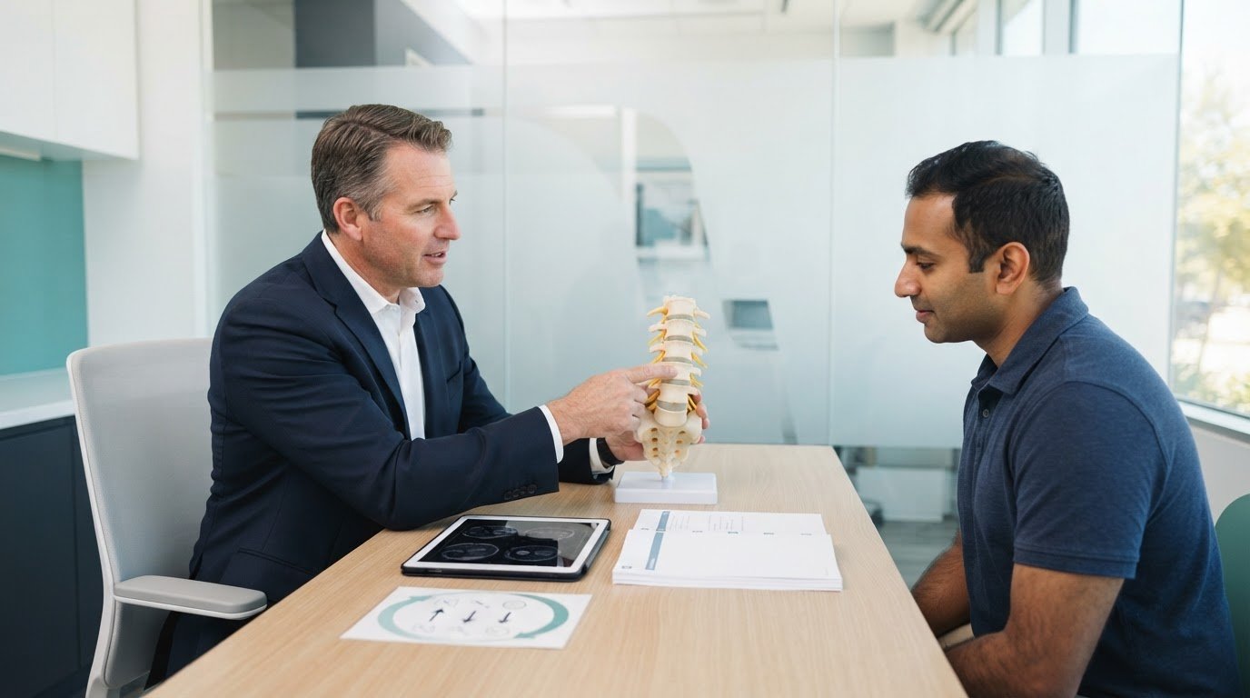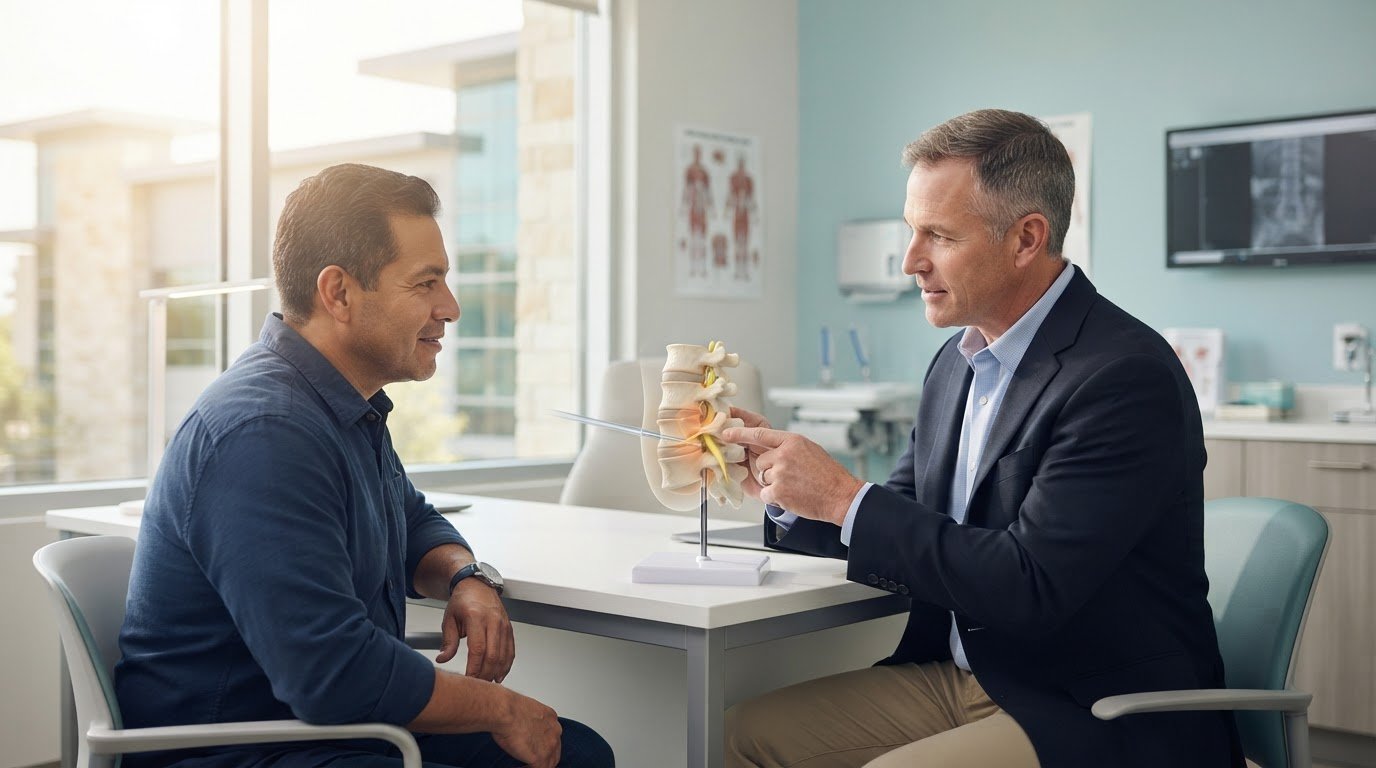
Minimally Invasive Neurosurgery of Texas provides endoscopic spine surgery in Lewisville and Plano, Texas. Led by Dr. Scott Kutz, the practice evaluates whether a small-incision procedure can relieve pressure on a compressed spinal nerve. A Lewisville endoscopic foraminotomy specialist considers the source of your symptoms and your spinal anatomy when discussing whether this approach may fit.
Back or leg pain, numbness, tingling, and weakness can make everyday movement difficult. Identifying the cause of these symptoms is an important first step toward choosing care suited to your condition.
This article explains how endoscopic foraminotomy works, which spinal problems it may address, and how minimally invasive techniques differ from open surgery. It also describes what guides surgical candidacy and what to expect when exploring treatment in Lewisville and Plano.
Key Takeaways
- Endoscopic foraminotomy widens the opening where a spinal nerve exits, which can relieve pressure from selected cases of stenosis.
- Symptoms such as back or leg pain, numbness, tingling, and weakness can help point to nerve compression, while imaging helps identify its source.
- Endoscopic spine surgery uses a small camera and instruments through an incision less than an inch long.
- Surgical suitability depends on the cause of nerve compression, response to nonsurgical care, and spinal anatomy.
- Learn how minimally invasive spine surgery in Lewisville and Plano may fit into a treatment plan for spinal conditions.
What is Endoscopic Spine Surgery?
Endoscopic spine surgery uses a small camera and instruments through a small incision to reach a specific spinal problem. Foraminotomy widens the opening where a spinal nerve exits the spine, relieving pressure caused by narrowing.
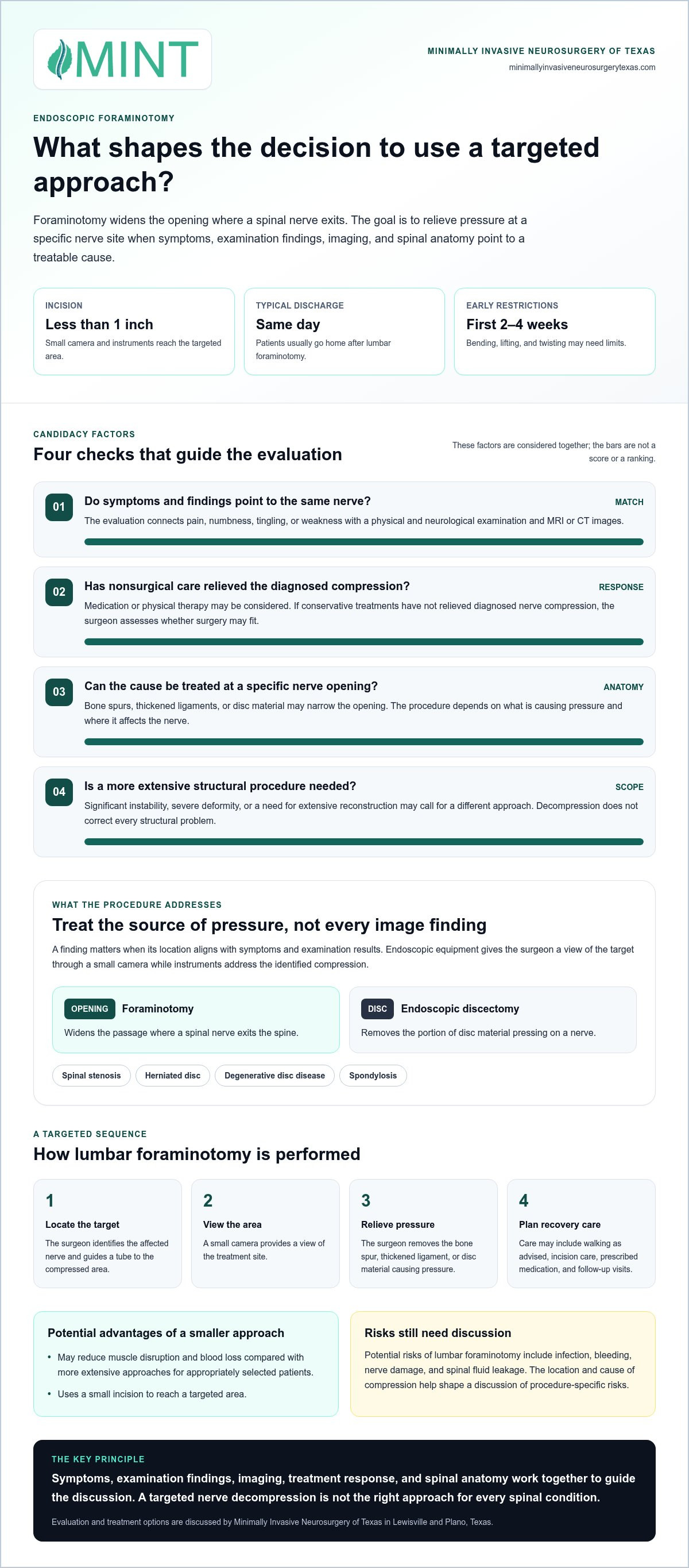
A spine surgeon first compares your symptoms with findings from a physical and neurological examination and MRI or CT imaging. If conservative treatments, such as medication or physical therapy, have not relieved diagnosed nerve compression, the surgeon considers whether your spinal anatomy is suited to an endoscopic approach.
Dr. Scott Kutz is a board-certified neurosurgeon and Fellow of the AANS and ACS. These credentials describe his professional qualifications, but they do not determine on their own whether a particular procedure suits your condition.
No specific Lewisville-area hospital or surgery center is identified here. The operating location depends on the surgeon’s treatment plan and the facility where the surgeon performs the procedure.
Insurance coverage varies by plan, diagnosis, and review requirements. Review your policy’s benefits and authorization rules to understand how your plan handles endoscopic foraminotomy.
Lumbar foraminotomy carries potential risks, including infection, bleeding, nerve damage, and spinal fluid leakage. The location and cause of nerve compression help shape a discussion of risks specific to the procedure.
Conditions Treated With Minimally Invasive Surgery
Minimally invasive spine surgery may address spinal stenosis, which can narrow the spinal canal or nerve openings and press on nerves, and herniated discs, where disc material can irritate or compress a nerve. Degenerative disc disease and spondylosis, age-related changes in spinal discs and joints, can also narrow nerve openings. Evaluation connects imaging findings with symptoms such as back or leg pain, numbness, and weakness.
Potential Benefits and Recovery
For appropriately selected patients, endoscopic surgery can reduce muscle disruption and blood loss compared with more extensive approaches. A small incision gives the surgeon access to the targeted area, but it does not remove the need to consider risks or follow recovery instructions. Recovery varies with the procedure and personal factors; patients may need to limit bending, lifting, and twisting for the first 2 to 4 weeks.
For lumbar foraminotomy, the surgeon identifies the target and guides a tube to the compressed nerve, then removes bone spurs, thickened ligaments, or disc material that is causing pressure. Patients usually go home the same day after lumbar foraminotomy. Recovery care may include regular walking as advised, incision care, prescribed medication, and follow-up visits.
Conditions treated, including stenosis and herniated discs
Endoscopic foraminotomy targets pressure on a spinal nerve where it exits between vertebrae. It may address foraminal stenosis, a narrowing of that nerve opening, when the compression matches your symptoms and imaging findings.
Symptoms can differ by spinal region. Lumbar nerve compression may cause lower back pain, leg pain, numbness, or weakness. Cervical nerve compression may cause neck pain or stiffness, with tingling, numbness, pain, or weakness extending into an arm or hand. These patterns help guide evaluation, but diagnosis must identify the actual source.
Several conditions can narrow the foramen, the passage through which a spinal nerve exits the spine:
- Spinal stenosis: Narrowing of a spinal passage can crowd nearby nerves.
- Herniated disc: Disc material can press against a nerve; an endoscopic discectomy removes the portion causing pressure.
- Degenerative disc disease: Changes in spinal discs can contribute to a narrowed nerve opening.
- Spondylosis: Degenerative changes in the spine can create pressure around nearby nerves.
Targeted decompression relieves pressure at the specific nerve site rather than treating every abnormality on an image. An evaluation connects pain, numbness, tingling, or weakness with physical findings and MRI or CT images. For example, a herniated disc on imaging is relevant when its location aligns with your nerve symptoms and examination.
Endoscopic equipment lets the surgeon view the target through a small camera and use instruments to address the compression. Foraminotomy widens the nerve opening; discectomy removes the disc portion pressing on the nerve. The procedure depends on whether bone, thickened ligament, or disc material is causing the narrowing. An accurate diagnosis helps identify the cause and match treatment to the affected spinal level.
Not every spinal condition is suited to endoscopic decompression. Significant spinal instability, severe deformity, or a need for extensive reconstruction may call for a different surgical approach. Conditions requiring fusion, a procedure that joins spinal bones, may also need another approach. Relieving pressure on one nerve does not correct every structural problem.
Endoscopic foraminotomy addresses symptoms linked to a treatable compressed nerve, not back or neck pain from every cause. Persistent leg symptoms associated with lumbar stenosis or arm symptoms associated with cervical nerve compression warrant evaluation of the nerve source. Diagnosis, symptom pattern, and spinal anatomy guide the treatment discussion and recovery plan.
These symptoms warrant evaluation for a specific compressed nerve. Degenerative disc disease, stenosis, herniated discs, and spondylosis can narrow the foramen and put pressure on nearby nerves. The procedure depends on the condition and the extent of the structural problem. Some spinal conditions, including those requiring fusion, may be better suited to a different surgical approach.
Incision size and surgical precision
Endoscopic spine surgery reaches a targeted area through an incision less than an inch long. Some procedures use incisions as small as 8 mm. The surgeon passes a small camera and instruments through the opening, viewing the spinal structures on a screen. This approach gives access to a specific nerve-compression site without the larger exposure used in traditional open surgery.
Before the procedure, the surgeon reviews your symptoms, performs a physical examination, and checks neurological function. This assessment evaluates strength, sensation, and other signs of nerve function. MRI or CT imaging helps locate the compression and show whether bone, thickened ligament, or disc material is pressing on the nerve. These findings guide the surgical target and plan.
For lumbar foraminotomy, the surgeon uses X-ray imaging to identify the surgical site. Fluoroscopy displays real-time X-ray images of the spine on a screen and helps guide instruments during surgery. You lie face-down under anesthesia while the surgeon makes a small incision over the target. A tube then guides instruments toward the compressed nerve.
Through the tube, the surgeon uses an endoscopic camera to view the treatment area and guide the procedure. The surgeon may remove a bone spur, thickened ligament, or part of a disc if it presses on the nerve. These structures can narrow the opening where the nerve exits. The goal is focused decompression, relieving pressure at the identified site.
Imaging and direct visualization have different roles. X-ray guidance helps locate the spinal level, while the endoscopic camera shows the working area. During lumbar foraminotomy, a microscope may also help the surgeon assess whether nerve decompression is complete. Together, these steps connect the pre-surgical diagnosis with the tissue causing compression.
After treating the target, the surgeon removes the instruments and guiding tube, then closes the incision. A small access point limits the size of the surgical opening, but incision size alone does not determine whether the procedure suits you. The surgeon considers your spinal anatomy, nerve symptoms, imaging findings, and the amount of decompression needed.
For example, a localized lumbar nerve compression may call for a different plan than compression at several levels or spinal instability. MRI or CT findings help define the problem before surgery. Precise targeting matters: the surgeon plans the incision and instrument path to reach the nerve requiring relief while protecting nearby spinal structures.
Minimally invasive and ultra-minimally invasive techniques
“Minimally invasive” describes how a surgeon reaches and treats a spinal problem. In endoscopic spine surgery, the surgeon uses a camera and instruments through a small incision to target nerve compression. “Ultra-minimally invasive” can describe an approach with limited access and tissue disruption, but the label alone does not identify the procedure. Ask what the surgeon plans to treat, how they will reach it, and which structures they intend to preserve.
The practical contrast is the route to the spine. The American Academy of Orthopaedic Surgeons describes a traditional open spine surgery incision as 5 to 6 inches long. Endoscopic techniques use a much smaller access point, letting the surgeon work through a narrow path rather than exposing a wider area. A smaller incision describes access, not the full complexity of the operation or the amount of nerve compression.
Muscle handling also matters. A minimally invasive cervical foraminotomy can avoid cutting and stripping muscles from the spinal region, reducing disruption to the tissues around the target. However, the approach must still provide enough access to remove material pressing on the nerve and assess the decompression.
For foraminal stenosis, which narrows the opening where a spinal nerve exits, the surgeon considers the location and cause of pressure. Bone spurs, thickened ligaments, or disc material may contribute. Instruments can widen the affected opening and remove the obstruction pressing on the nerve. The appropriate technique depends on your anatomy and the planned extent of treatment.
Not every diagnosis can be treated through a narrow endoscopic route. Significant spinal instability, severe deformity, or a need for extensive reconstruction may call for another surgical approach. Conditions requiring fusion, which joins spinal bones, may also need a different plan. A smaller incision cannot correct instability by itself, so the surgeon must match the technique to the structural problem.
During a consultation, focus on the planned target and why the surgeon recommends a particular access method. Ask how imaging relates to your symptoms, which tissue is causing compression, and whether the proposed procedure addresses that cause. These details help distinguish a specific endoscopic plan from a broad label such as “ultra-minimally invasive.”
The central takeaway: compare the procedure, not the label. For someone with localized nerve compression and suitable anatomy, endoscopic techniques may provide focused access with less muscle disruption. If reconstruction or correction of instability is needed, the surgeon must consider a broader approach. The diagnosis, anatomy, and surgical goal determine which technique makes clinical sense.
Which spine surgeons in or near Lewisville, Texas specialize in endoscopic foraminotomy?
Dr. Scott Kutz is a neurosurgeon serving the Lewisville area who evaluates spinal conditions and surgical options. He is board-certified in neurosurgery and a Fellow of the AANS and ACS. These credentials describe his specialty and professional affiliations. A procedure-specific consultation determines whether endoscopic foraminotomy suits your diagnosis and spinal anatomy.
When seeking a Lewisville endoscopic foraminotomy specialist, focus on how the surgeon’s training relates to the condition requiring treatment. Ask how the surgeon identifies the compressed nerve, interprets MRI or CT images, and decides whether focused decompression can address the cause. A clear explanation should connect your symptoms and imaging findings to the proposed surgical target.
Dr. Kutz serves patients in Lewisville and Plano, Texas. During an evaluation, the surgeon reviews your history of back, leg, neck, or arm symptoms, performs a physical and neurological examination, and considers imaging. Neurological findings may include changes in strength or sensation. These steps help distinguish a nerve-root problem from pain caused by another spinal condition.
Useful points to discuss during a surgical consultation include:
- Diagnosis: Which finding explains the patient’s symptoms, such as foraminal stenosis or a herniated disc?
- Procedure: What tissue creates pressure, and how would the planned operation address it?
- Approach: Does the spinal anatomy suit endoscopic access, or does instability or deformity change the plan?
- Recovery: Which activity limits and follow-up steps apply after the proposed procedure?
These questions focus the discussion on clinical reasoning, not broad labels such as “minimally invasive.” For example, the surgeon should explain whether bone, thickened ligament, or disc material narrows the nerve opening. The explanation should also clarify how the surgeon plans to relieve compression while accounting for nearby nerves and spinal structures.
Board certification and fellowship affiliations provide important background, but the treatment plan must address your individual diagnosis. Localized nerve compression may call for a different approach than severe deformity, instability, or a need for extensive reconstruction. These structural findings can affect which surgical approach is appropriate.
Patients in Lewisville and Plano can arrange an evaluation with Dr. Scott Kutz to discuss persistent nerve-related symptoms and surgical options. Bring relevant MRI or CT images and a concise record of your symptoms, including where pain travels and whether numbness or weakness affects daily activities. These details help focus the consultation on diagnosis, anatomy, and a suitable next step.
Endoscopic spine surgery may be an option for selected patients with foraminal stenosis. Patients usually return home the same day after endoscopic spine surgery.
Minimally Invasive Neurosurgery of Texas: Minimally Invasive Spine Surgery
Minimally Invasive Neurosurgery of Texas provides spinal care in Lewisville and Plano, Texas, including endoscopic spine surgery with lead surgeon Dr. Scott Kutz. Patients with persistent back or leg symptoms can discuss whether a focused surgical approach fits their diagnosis and daily needs.
Dr. Kutz is a board-certified neurosurgeon and Fellow of the AANS and ACS. Evaluation connects your symptoms, imaging, and spinal anatomy with the treatment options under consideration.
If ongoing symptoms affect your daily activities, contact Minimally Invasive Neurosurgery of Texas to discuss an evaluation and possible next steps.
Take the Next Step Toward a Clear Treatment Plan
Start by noting what your symptoms interrupt, such as walking, sleep, or work. Bring that information and your imaging to a consultation so the discussion can connect daily challenges with your spinal diagnosis and possible treatment goals.
A Lewisville endoscopic foraminotomy specialist can assess whether the location and cause of nerve compression suit a focused procedure. Minimally Invasive Neurosurgery of Texas offers minimally invasive neurosurgery, minimally invasive spine surgery, and artificial disc replacement. The appropriate treatment depends on your condition, spinal anatomy, and goals of care.
Dr. Scott Kutz can discuss your diagnosis and surgical options in a clear, patient-focused conversation. Contact Minimally Invasive Neurosurgery of Texas to take the next step toward understanding your options and planning care with confidence.
Frequently Asked Questions
How is the procedure performed?
The surgeon’s plan identifies the spinal level, side, and nerve linked to your symptoms. The procedure targets structures contributing to that specific compression rather than every change visible on imaging. Before proceeding, ask which finding guides the plan and what the surgeon would do if your anatomy differs from the images. This discussion clarifies the intended scope of the procedure.
What Happens During the Lumbar Foraminotomy Procedure?
During a lumbar procedure, the surgeon treats the lower-back nerve exit identified during your evaluation. The surgical plan specifies the level and amount of decompression needed. The surgeon focuses on relieving pressure at that target, rather than treating unrelated spinal changes. Before surgery, discuss how the care team will assess your symptoms afterward and which instructions will guide your first steps in recovery.
What is a Foraminotomy?
A foraminotomy creates more space for a spinal nerve where it leaves the spine. It focuses on the nerve’s exit route rather than removing an entire disc or joining spinal bones. That distinction matters because a herniated disc, a narrowed nerve opening, and spinal instability are different problems. Diagnosis determines whether widening the nerve passage addresses the cause of your symptoms.
Who requires this procedure?
A specialist may consider this procedure when nerve compression matches your symptoms and affects everyday function. A symptom alone, such as leg pain or tingling in the hand, does not establish that foraminotomy is appropriate. Evaluation considers your symptom pattern, examination findings, and imaging together. Share which movements or daily tasks worsen your symptoms to help explain their practical impact.
How does endoscopic foraminotomy compare with open or minimally invasive foraminotomy?
Endoscopic foraminotomy uses a camera-guided route, while open and other minimally invasive approaches use different ways to access the spine. The surgeon considers how much access the target requires and whether the operation must address problems beyond nerve compression. Ask what the proposed method allows the surgeon to treat and why it suits your anatomy. The approach should match the surgical goal, not just a label.
What are the success rates and recovery time for endoscopic foraminotomy?
No single success rate or recovery schedule applies to every endoscopic foraminotomy. Outcomes depend in part on whether the treated nerve explains the symptoms and whether the procedure addresses that source. Recovery also reflects the extent of the operation and individual health factors. Ask how your care team will track changes in pain, sensation, strength, and daily function, and which symptoms should prompt follow-up.
How can patients prepare for an endoscopic foraminotomy consultation and surgery?
Prepare for the consultation by bringing your imaging, medication and allergy lists, and records of prior spine procedures. Note when symptoms began, where they travel, and which activities they disrupt. Ask the surgeon to explain the diagnosis, treatment goal, alternatives, and recovery instructions. Before surgery, follow the care team’s directions for preparation and medication use rather than changing medicines on your own.