
Minimally Invasive Neurosurgery of Texas provides minimally invasive spine surgery in Lewisville and Plano, Texas. Dr. Scott Kutz is a board-certified neurosurgeon specializing in spinal care. Learning how to read your own spine MRI report can help you understand the findings and prepare focused questions for a discussion about your care.
It’s understandable to focus on terms such as “bulge” or “herniation,” especially when a newer report sounds more serious. These words describe what appears in the images, but they don’t determine by themselves whether a finding explains your pain or means surgery is appropriate.
This guide explains how to compare cervical MRI findings, understand sagittal views (side-on images) and axial views (cross-sections), and interpret notes about alignment and discs. It also covers how protrusions differ from extrusions, and why those details need to match your symptoms before they inform treatment decisions.
Key Takeaways
- Compare reports level by level, and distinguish a real imaging change from wording that simply differs between radiologists.
- Use T1- and T2-weighted images to understand how fat and fluid appear, while treating signal changes as clues rather than diagnoses.
- Read sagittal, axial, and coronal views together to assess alignment, disc shape, and possible nerve contact.
- Disc bulges, protrusions, and extrusions describe different appearances, but their importance depends on location and how well they match your symptoms.
- MRI findings alone don’t determine whether surgery makes sense; symptom patterns, examination findings, and the images all inform the decision.
Differences between T1- and T2-weighted images
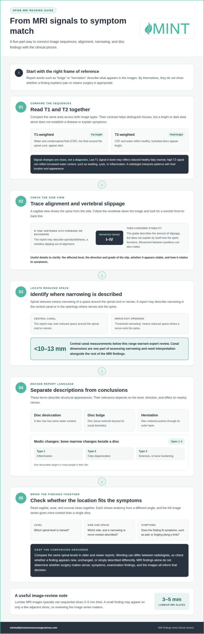
A spine MRI uses radio waves and powerful magnets to create detailed images without radiation. Radiologists use different image sequences to emphasize different tissues. On T1-weighted images, fat appears bright, while water and cerebrospinal fluid (CSF), the fluid around the spinal cord, appear dark. On T2-weighted images, fluid appears bright, including CSF and the water within healthy, hydrated discs. These contrasts help clinicians inspect anatomy and identify possible changes.
On a lumbar MRI disk, you’ll typically find two basic image types: T1-weighted and T2-weighted. Compare the same area across both sequences rather than drawing conclusions from one bright or dark spot. For example, a fluid-filled structure may appear dark on T1 and bright on T2. That contrast can help clinicians distinguish fluid from surrounding tissue, but brightness alone doesn’t establish a disease or explain symptoms.
Signal changes provide clues, not a diagnosis. Low T1 signal in bone may reflect reduced healthy fatty marrow and may occur with infection, trauma, or lesions, meaning tissue abnormalities. High T2 signal can reflect increased water content, such as swelling, cysts, or inflammation. A radiologist interprets those patterns alongside the structure’s location and appearance. The words “normal” or “severe” in a report also need context, especially when it describes narrowing, or reduced space around nerves.
To obtain your spine MRI images and report in the United States, contact the imaging facility’s medical records or radiology department. Request both the written report and the image files, and bring them to your appointment.
Ask your doctor which finding matches your symptoms, what the T1 and T2 sequences show, and whether any narrowing or signal change needs follow-up. For example, ask whether a bright T2 area represents fluid or another tissue change.
MRI slices show the body in thin sections, not as one continuous photograph. Lumbar MRI images typically use sequential slices 3-5 mm thick. A small finding may appear on only a few adjacent slices, so the reader must follow its position through the image series. Compare matching spinal levels and note whether a finding appears on T1, T2, or both.
Bring the actual images, not just a printed report or list of findings. A clinician can review the image series and compare a reported change with nearby anatomy. Keep the date and body region of each study clear, such as “cervical MRI” for the neck, so older and newer examinations aren’t confused.
Reviewing the sequences can help you ask focused questions, but it doesn’t replace clinical interpretation. A thorough review connects the report’s wording with the images, the affected spinal level, and your symptoms.
Spinal alignment and vertebral slippage
A sagittal MRI view shows the spine from the side, making it useful for assessing vertebral alignment and disc height. Follow the vertebrae down the image and look for a smooth front-to-back line. If one vertebra sits farther forward or backward than its neighbor, the report may describe spondylolisthesis, a slip out of alignment. It may also note the direction and grade of the slip.
Spondylolisthesis is graded from I to IV. The grade describes the amount of slippage, but doesn’t explain by itself how the spine functions. Stability matters too: movement between positions can affect whether a slip causes symptoms or influences treatment planning. Ask which vertebral level is affected, whether the slip appears stable, and how it relates to your symptoms.
Spinal stenosis means narrowing of a space around the spinal cord or nerves. Spinal stenosis can involve narrowing in the central canal or in the openings where nerves exit the spine. A report may also use “foraminal narrowing,” meaning reduced space in that nerve exit. Central canal measurements below 10-13 mm warrant expert review. Canal dimensions are one part of assessing spinal narrowing and should be interpreted alongside the rest of the MRI findings.
Common degenerative findings describe structural changes that develop over time. “Disc desiccation” means a disc has lost some water content, while “disc bulge” describes disc tissue extending beyond its usual boundary. “Herniation” means disc material pushes through its outer layer. These terms describe appearances, not a complete diagnosis. The level, direction, and effect on nearby nerves help clarify their relevance.
Disc desiccation begins in many people in their 30s. Modic changes describe bone marrow changes beside a disc: Type 1 indicates inflammation, Type 2 fatty degeneration, and Type 3 sclerosis, or bone hardening. Those labels help characterize pathology, meaning a tissue change or disease process. Compare the specific levels and descriptions in old and new reports to see whether a finding appears new, unchanged, or different.
For a useful comparison, note the level, side, and any reported narrowing or slippage. Then relate those details to symptoms, such as pain or tingling along a particular limb. An MRI finding is most meaningful when its location and effects fit the clinical picture.
Axial, coronal, and sagittal MRI views
Spine MRI images show anatomy from three angles: sagittal, axial, and coronal. Each view highlights different relationships between vertebrae, discs, nerves, and surrounding tissue. Reviewing them together helps show where a finding is and whether it affects nearby structures. One image is only a slice through the body, so the full series provides more context.
A sagittal view shows the spine from the side. It helps readers follow the spinal curve, compare disc spaces, and locate findings by level. For example, a cervical sagittal image can show whether a disc projects toward the spinal canal. The lumbar mid-sagittal region extends 7 mm on either side of the true mid-sagittal cut. Comparing neighboring sagittal slices helps distinguish a central finding from one positioned farther to the side.
An axial view is a horizontal cross-section through the body. In a lumbar MRI, it can show the vertebral body in front, the spinal canal behind it, and nerve roots extending toward each side. This view can help show whether a disc is pressing on a nerve root and lets the reader compare the left and right sides at the same level. For example, a report may describe more pressure near one nerve exit than the other.
A coronal view shows the spine from the front. It helps compare left-to-right alignment and may make uneven positioning or an off-center finding easier to see. This perspective can add context when a sagittal image suggests a shift and an axial image shows how surrounding tissue relates to the spinal canal. No single view shows every feature equally well.
Check the labels on each image series to identify the view and spinal level before comparing findings. Match the same level across sagittal and axial images, then use coronal images to assess alignment from the front. For example, if an axial slice shows narrowing near a nerve root, the sagittal series can help locate that slice along the spine. Comparing views connects the report’s wording with the anatomy.
The lumbar spine has five vertebral bodies and five lumbar discs. MRI images show these structures in successive slices, allowing clinicians to follow a finding across nearby tissue. A disc can look different at its center than near its side, so location matters. The three views can help you understand what the report describes, while the radiologist’s written interpretation organizes the images into findings.
The Big Question: Do I Need Surgery Based on My Cervical Spine MRI Results?
No. Cervical MRI results alone don’t determine whether surgery is appropriate. The decision depends on how the images relate to your symptoms, examination findings, and the specific spinal level.
Matching Findings to Symptoms
Clinical correlation means matching MRI findings with your symptoms and physical examination. This matters because an image can show a disc change without explaining your pain, numbness, or weakness.
Compare the finding’s level and side with where your symptoms travel. For example, a cervical disc pressing on a nerve may correspond to arm pain or tingling on the same side. A finding that doesn’t match your symptoms may have a different significance. At a consultation, ask which MRI finding best explains your symptoms and what examination results support that connection.
Incidental Findings Without Pain
An incidental finding is an MRI change that appears in someone without related pain. A visible abnormality doesn’t automatically mean disease or a need for treatment.
A study found abnormalities on 19% of cervical MRI scans among people without symptoms. Among asymptomatic people over 40, 28% had abnormalities. Disc changes also appear in people without back pain: approximately 30% of adults without low back pain have a disc protrusion. These examples show why a report must be read alongside symptoms, not as a diagnosis on its own.
Indication, Findings, and Impression
The indication states the symptoms or medical reason for the MRI. The findings section records the radiologist’s observations, including normal anatomy, abnormal changes, and age-related features.
The impression summarizes the key findings and may suggest a diagnosis for the treating clinician to review. Read the indication first to understand the question behind the scan, then compare it with the impression. Use the findings section to locate details supporting the summary. For example, an impression describing nerve compression should correspond to a specific level and observation in the findings.
Findings Warranting Clinical Attention
Findings warrant clinical attention when they could affect the spinal cord, nerve roots, or surrounding structures. Central canal stenosis below 10-13 mm warrants expert review. Stenosis means narrowing; the report’s location and degree help explain its significance.
Bone marrow edema means swelling from fluid buildup and can follow injury or severe degeneration. A new or changing finding deserves comparison with earlier images, especially if symptoms have also changed. An unchanged cervical disc herniation present 5 years earlier is unlikely to explain new pain unless it has grown or calcified. During consultation, ask what changed, where it appears, and whether it matches your symptoms.
Disc bulges, herniations, and extrusions
Disc terms describe different shapes, not a scale from harmless to dangerous. A bulge extends beyond the disc’s usual border across a broader area. A herniation describes disc material pushing through its outer layer in a more localized area. An extrusion is a herniation in which the material extends farther through that layer. These distinctions describe what the radiologist sees, but shape alone doesn’t identify the main cause of pain.
A protrusion is a small disc herniation measuring less than 4-5 mm. Its size matters less than its position and nearby anatomy. For example, a modest protrusion beside a nerve root, the nerve branch leaving the spine, may relate to symptoms if it presses on that nerve. A larger bulge that doesn’t contact a nerve may have a different clinical significance.
Read the level and direction alongside the shape. A report might describe a right-sided herniation at a cervical level, then note whether it narrows the spinal canal or a nerve exit. “Central” means toward the middle; “right” or “left” identifies the side. Compare the same level and side in older and newer reports. A change in wording may reflect a more specific description, while comparing images can show whether the disc shape or nerve contact actually changed.
Look for the radiologist’s description of nerve root contact, displacement, or compression. Contact means the disc touches the nerve; displacement means it moves the nerve from its usual position; compression means it presses on the nerve. These terms describe different relationships. Match the affected side and level with symptoms, such as pain or tingling that travels into an arm or leg. The most dramatic phrase in a report may not describe the finding that best matches your symptoms.
Disc bulges appear in over half of adults without symptoms. This makes context essential: a bulge can be present without causing pain. Degenerative disc disease describes age-related changes in disc structure, but the term alone doesn’t establish that a disc causes someone’s symptoms. The location, nearby nerves, and clinical picture help put the finding in context.
For a useful comparison, note each report’s level, side, disc shape, and effect on nearby nerves. Then compare those descriptions with the images and your symptom pattern. If a newer report describes an extrusion, ask whether it changes the nerve relationship or simply uses more specific wording. This can help prevent a scan finding from becoming a treatment decision by itself.
Minimally Invasive Neurosurgery of Texas: Minimally Invasive Spine Surgery
Minimally Invasive Neurosurgery of Texas provides minimally invasive spine surgery in Lewisville and Plano, TX. Dr. Scott Kutz, a board-certified neurosurgeon and Fellow of the AANS and ACS, emphasizes that MRI findings need to fit the patient’s symptoms and examination. For patients considering surgical care, minimally invasive spine techniques offer an option to discuss in that context.
Patients can learn more about the practice’s approach through Minimally Invasive Neurosurgery of Texas and consider whether a conversation about their MRI would help clarify next steps.
If you’re ready to discuss your MRI, contact Minimally Invasive Neurosurgery of Texas.
Turn Your MRI Review Into a Clear Next Step
Use your MRI review to prepare a focused conversation about what matters in your daily life. Before a consultation, write down which activities have become difficult, how your symptoms affect them, and what you hope to regain. These priorities help keep treatment discussions centered on your goals, not just the wording in a report.
Learning how to read your own spine MRI report can help you arrive with specific questions, but you don’t need to interpret every image alone. A specialist can explain how the scan, symptoms, and examination fit together and discuss care options for your situation. This conversation can clarify whether monitoring, nonsurgical care, or a surgical approach warrants consideration.
If you’re ready to talk through your MRI and next steps, contact Minimally Invasive Neurosurgery of Texas. A thoughtful conversation can help you move forward with greater clarity and confidence.
Frequently Asked Questions
How do you compare old and new cervical MRI reports to assess progression?
Start by noting whether your symptoms changed between scan dates, including where they occur and which activities they affect. Then ask the clinician whether the newer study changes the care plan. Different wording in a report may not represent meaningful progression. A useful comparison connects any imaging change with changes in symptoms or function.
Should I get a second opinion on cervical MRI interpretation?
A second opinion is reasonable before major spine surgery. Use the discussion to clarify the proposed goal: which symptom the procedure aims to address, which MRI finding supports that choice, and what alternatives the clinician considered. Ask how the expected benefit relates to your daily limitations. These questions can help you understand the reasoning behind a recommendation rather than focusing only on the report’s most concerning phrase.
What Does a Spine MRI Show?
A spine MRI can help clinicians assess discs, nerves, the spinal cord, and other soft tissues. For many patients with neck pain or radiculopathy, meaning symptoms caused by an irritated nerve, cervical MRI without contrast can identify disc herniations and stenosis. The clinician uses the scan to evaluate a specific concern, such as pain traveling from the neck into an arm.
What sections are included in a spine MRI report?
Reports may also describe the imaging technique and comparisons with earlier studies. These details explain how the radiologist produced the images and whether the report refers to prior scans. If you see a “comparison” note, check which earlier examination it names. This can clarify whether the radiologist directly compared earlier images or reviewed only the current study.
What do cervical, thoracic, and lumbar spine MRI findings mean?
The region named in the report tells you which part of the spine the findings concern. Cervical refers to the neck, thoracic to the middle back, and lumbar to the lower back. This distinction helps avoid confusion when symptoms involve more than one area. For example, a lumbar report doesn’t describe the neck, even if you have symptoms in both regions.
How to interpret levels and locations in a spine MRI report?
“Multilevel” means the report describes findings at more than one spinal level. “Bilateral” means both sides, while “unilateral” means one side. These terms help describe the extent and distribution of a finding. If you see an unfamiliar term, ask the clinician to point to the relevant area on the images and explain its practical significance.
What is the difference between common age-related MRI findings and clinically significant problems?
A structural change matters in context, not simply because it appears on a scan. Consider whether the finding relates to a specific limitation, such as difficulty turning your head or completing a usual activity. A report may describe a change without explaining its effect on daily function. Discuss your symptoms and examination, not just the descriptive wording, when reviewing its significance.
How do MRI findings correlate with symptoms and physical examination?
A physical examination can assess functions that an MRI image cannot measure directly. For example, a clinician may check arm strength, sensation, grip, and reflexes alongside a neck MRI. The pattern across these checks can help determine whether a scan finding fits your experience. This content is for informational purposes only, isn’t a substitute for professional medical advice, and the website is provided as-is.