By the age of 65, 95% of people in the U.S. will show evidence of cervical spondylosis, yet most only learn about their cervical vertebrae when radiating pain begins to interfere with their daily work. You might be staring at an MRI report filled with intimidating jargon while worrying that an invasive open neck surgery is your only path to relief. It’s completely normal to feel overwhelmed by the complexity of your spine and the high stakes of surgical intervention.
We’re here to provide a clear roadmap for your recovery. We promise to help you unlock a clear understanding of your neck’s structure and show you how advanced, minimally invasive treatments can restore your quality of life. This guide covers the seven levels of the neck, identifies common causes of pain, and explores modern, low-impact surgical options like artificial disc replacement. You’ll discover how our focus on precision technology and motion preservation can help you return to normal activity with confidence.
Key Takeaways
- Understand the critical function of the seven cervical vertebrae and how they balance structural support with spinal cord protection.
- Recognize the hallmarks of radiculopathy and how nerve compression from herniated discs or stenosis impacts your daily physical function.
- Learn why advanced diagnostic imaging is essential for distinguishing between various cervical disorders and guiding effective treatment.
- Evaluate modern, low-impact surgical options like artificial disc replacement that prioritize motion preservation and long-term spinal health.
- Discover how specialized minimally invasive neurosurgery can significantly reduce recovery times and help you achieve a rapid return to normal activity.
The Foundation of Neck Health: What Are Cervical Vertebrae?
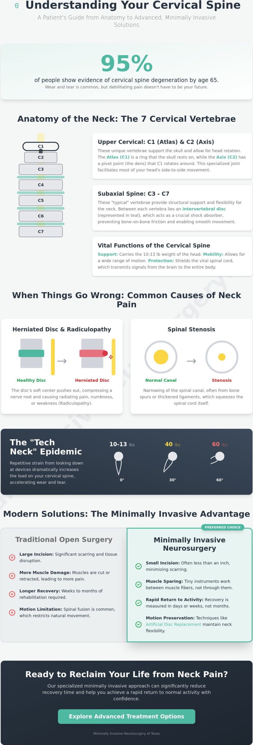
The human neck is a structural marvel, composed of seven distinct bony segments known as the cervical vertebrae. Labeled C1 through C7, these bones form the uppermost portion of the spinal column. While they’re the smallest vertebrae in your body, their responsibility is immense. They must support the weight of your head, which typically weighs between 10 and 13 pounds, while providing a protective “tunnel” called the vertebral foramen for your spinal cord. This delicate balance of strength and space is what allows you to move freely without compromising your neurological health. Unlike the rigid thoracic spine, which is anchored by the rib cage, the cervical region is designed for extreme mobility.
The Vital Functions of Your Cervical Spine
Your cervical spine serves three primary purposes that dictate your daily quality of life. First, it provides the structural scaffolding necessary to maintain an upright posture. Second, the cervical region offers a range of motion far exceeding other spinal areas. It facilitates complex movements like rotation, flexion, and extension, allowing you to scan your environment with ease. Finally, it acts as a high-security gateway. Every signal sent from your brain to your limbs must pass through this narrow corridor, making the health of these cervical vertebrae essential for total body communication.
- Structural Support: The vertebrae stack precisely to maintain balance and distribute the weight of the skull.
- Range of Motion: Specialized joints allow for the tilting and turning of the head.
- Neurological Protection: The spinal canal houses the cord, protecting it from external trauma.
Why the Neck is Vulnerable to Injury
The very flexibility that makes the neck useful also makes it prone to degeneration. Because the cervical vertebrae are highly mobile and carry significant weight, they experience consistent mechanical stress. In our local communities across Plano and Lewisville, we’re seeing an increase in “tech neck.” This condition is caused by the repetitive postural strain of looking down at mobile devices, which can double or even triple the effective weight on your spine. This modern habit, combined with the natural aging process, often leads to reduced disc hydration and shifting bone density. By age 65, approximately 95% of Americans show evidence of cervical spondylosis, highlighting how common wear and tear is in this region. When these structures wear down, the resulting pain can be debilitating, often requiring a specialized neurosurgical evaluation to determine the best path forward for recovery.
Anatomy of the Cervical Spine: C1 through C7 Explained
The cervical region is far more than a simple stack of bones. It’s a sophisticated architectural unit divided into three distinct functional levels: the upper, middle, and lower cervical spine. Each of these segments houses specific cervical vertebrae with specialized shapes that dictate how you move and how your nervous system is protected. A detailed look at the Anatomy of the Cervical Spine reveals a structure designed for both immense protection and incredible range of motion. Understanding this layout is the first step toward demystifying your MRI results and choosing the right treatment path. The cervical spine anatomy is a complex system of bone, nerve, and disc.
The Upper Cervical Level: Atlas (C1) and Axis (C2)
The first two bones in your neck are known as “atypical” because they don’t look like any other vertebrae in the spine. The Atlas, or C1, is a delicate, ring-shaped bone that serves as the “globe” supporting your skull. It doesn’t have a traditional vertebral body. Instead, it sits atop the Axis (C2). The Axis features a unique bony projection called the dens, which acts as a pivot point. This relationship allows for the majority of your head’s rotation. Because these levels sit so close to the brainstem, injuries or instability here are considered critical neurological events that require immediate, expert intervention to prevent long-term damage.
The Subaxial Spine: C3 through C7
Moving down the neck, the vertebrae from C3 to C6 are considered “typical.” They share similar characteristics, including small vertebral bodies and transverse foramina that allow vertebral arteries to pass through. Between these levels, intervertebral discs act as vital shock absorbers. They prevent bone-on-bone contact and provide the cushioning needed for fluid motion. If you’re wondering how many cervical vertebrae are there, the answer is seven, ending with the unique C7.
The C7 vertebra, also called the Vertebra Prominens, marks the transition to the thoracic spine. You can often feel this bone as a prominent bump at the base of your neck. It serves as a major attachment point for muscles that support your head and shoulders. When wear and tear affects these lower levels, it often leads to the radiating pain many patients describe as a “pinched nerve.” If you’re experiencing this type of discomfort, a consultation for minimally invasive spine surgery can help determine if your anatomy is contributing to nerve compression. Our team focuses on precise diagnostics to ensure we address the specific level of your pain with the least invasive methods possible.
When Cervical Vertebrae Fail: Common Conditions and Symptoms
When the structural integrity of your cervical vertebrae begins to decline, the impact is rarely limited to the neck alone. Many patients arrive at our clinic with significant anxiety, fearing that a diagnosis of spinal degeneration automatically leads to a high-risk, open neck surgery. This is a common misconception. Modern neurosurgery has evolved. Today, most conditions affecting the neck can be managed through advanced, low-impact techniques that prioritize your comfort and long-term mobility. Understanding the source of your pain is the first step toward relief.
Chronic pain and loss of flexibility often stem from the gradual breakdown of the components that support your head. As the discs between your vertebrae lose hydration or the bones themselves develop irregularities, the space available for your nerves begins to shrink. This narrowing is the root cause of radiculopathy. This is the clinical term for the radiating pain, numbness, or weakness that travels from your neck down into your shoulders and arms. It’s a signal that your anatomy is no longer providing the protection your nervous system requires.
Herniated Discs and Foraminal Stenosis
A herniated disc occurs when the soft inner material of a spinal disc pushes through a tear in the tougher outer layer. This protrusion often presses directly against the nerves exiting the cervical vertebrae. We distinguish between two types of narrowing: central stenosis and foraminal stenosis. Central stenosis involves the main spinal canal, potentially affecting the spinal cord itself. Foraminal stenosis occurs when the small openings where individual nerve roots exit become constricted. You might experience sharp, electric-shock sensations or a persistent tingling in your fingers. These symptoms are clear signals from your body that a nerve is under duress and needs professional evaluation.
Cervical Spondylosis and Bone Spurs
Cervical spondylosis is the medical term for age-related wear and tear. Over time, the body may attempt to stabilize a weakening spine by growing osteophytes, or bone spurs. While these are a natural response to instability, they often encroach on the neural pathways. This process can lead to a stiff neck and a reduced range of motion that makes simple tasks like driving or typing difficult. Identifying these changes early is vital. Timely diagnosis through high-resolution imaging allows us to intervene before permanent nerve damage occurs. Our goal is always to address these structural failures with precision, facilitating a swift return to normal activity through minimally invasive spine surgery.
Modern Diagnostic and Treatment Pathways for Cervical Disorders
Precision is the hallmark of modern spinal care. We no longer rely on general assessments. Instead, we utilize high-resolution imaging to create a digital map of your cervical vertebrae and the surrounding neural structures. The AO Spine Clinical Practice Recommendations (January 2026) now designate CT and MRI as the gold standard for radiological workup. These tools allow us to see exactly where bone spurs or herniated discs are encroaching on neural pathways. This clarity ensures that every intervention is targeted and necessary, moving you closer to a successful clinical outcome.
Many patients begin their journey with conservative management. Options such as activity modification, targeted injections, and physical therapy can be effective for mild symptoms. However, if these methods fail to provide lasting relief, we transition to structural correction. In 2026, the clinical focus has moved away from simply “managing pain.” We now look to restore the natural alignment and function of the spine through advanced surgical interventions that address the root cause of your discomfort.
Artificial Disc Replacement vs. Spinal Fusion
When surgery is required, the choice often comes down to Artificial Disc Replacement (ADR) or spinal fusion. ADR is a motion-preserving technology that mimics the natural function of the intervertebral discs between the cervical vertebrae. By maintaining flexibility, we can significantly reduce the risk of “adjacent segment disease,” where the levels above and below a fusion experience accelerated wear. For younger or more active individuals, Neck Disk Replacement in Plano & Lewisville is often the preferred choice to ensure a long-term return to normal activity. Fusion remains a vital tool when stability is the primary concern, such as in cases of severe spinal instability or deformity.
The Role of Minimally Invasive Techniques
Our practice specializes in Minimally Invasive Neurosurgery. This approach utilizes specialized instruments and high-tech imaging to access the spine through tiny incisions. This method dramatically reduces damage to the delicate muscles and ligaments surrounding the neck. Patients benefit from lower infection rates, reduced post-operative pain, and faster recovery timelines. Many of these procedures are now performed in an outpatient setting, allowing you to return home the same day. This high-tech approach ensures that your recovery is as efficient as the surgery itself.
If you’re ready to move beyond temporary fixes and address the root cause of your neck pain, contact our team to discuss minimally invasive spine surgery options that can restore your quality of life.
Minimally Invasive Solutions for Cervical Spine Health in North Texas
Minimally Invasive Neurosurgery of Texas (MINT) stands as the premier destination for advanced spine care in Plano and Lewisville. Led by Dr. Scott Kutz, a board-certified neurosurgeon, our practice focuses on correcting structural issues within the cervical vertebrae using the most sophisticated techniques available today. We recognize that chronic neck pain is more than a physical ailment; it’s a significant burden on your daily productivity and emotional well-being. Our mission is to provide the relief you need through specialized care that prioritizes your long-term mobility and comfort. Precision matters. We don’t just treat symptoms; we restore function.
Advanced Technology at MINT
Our commitment to excellence is reflected in our investment in cutting-edge surgical tools. We utilize the Globus ExcelsiusGPS robotic system to achieve unparalleled accuracy during every procedure. This proprietary technology allows Dr. Kutz to map the unique anatomy of your neck with sub-millimeter precision before the first incision is made. When combined with Augmented Reality (AR) systems, we can visualize the complex pathways around the cervical vertebrae in real-time. This high-tech approach ensures that your spine surgery is as safe and effective as possible. By minimizing disruption to healthy tissue, we facilitate a faster recovery and a more predictable clinical outcome.
Your Journey to Relief in Plano and Lewisville
Your path to recovery begins with a detailed consultation at one of our North Texas locations. Our team takes the time to listen to your history and perform a thorough neurological exam. We believe an informed patient is a confident patient. We explain your diagnosis in clear language, moving past the jargon of MRI reports to focus on actionable solutions. Residents of the Dallas-Fort Worth metroplex benefit from our specialized, boutique environment that avoids the coldness of large hospital systems. If you’re struggling with radiculopathy or persistent stiffness, don’t wait for the damage to become permanent. We’re here to help you achieve a rapid return to normal activity through minimally invasive neurosurgery at Minimally Invasive Neurosurgery of Texas. You deserve a life free from the constraints of chronic neck pain.
Take the Next Step Toward Lasting Neck Relief
Your journey through the complexities of the cervical vertebrae shouldn’t end with a diagnosis; it should end with a structural solution. By understanding the unique anatomy of your neck and the modern diagnostic pathways available, you’ve already taken the first step toward reclaiming your quality of life. We’ve explored how conditions like stenosis and herniated discs impact your mobility and why motion-preserving technologies like artificial disc replacement are now the clinical standard for active patients.
At Minimally Invasive Neurosurgery of Texas, we combine the expertise of board-certified neurosurgeon Dr. Scott Kutz with advanced robotic technology to ensure every procedure is performed with absolute precision. We’re dedicated to serving the Plano and Lewisville communities with a patient-centered approach that prioritizes a rapid return to normal activity. You don’t have to live with the limitations of chronic neck pain or the fear of traditional, invasive surgery.
Schedule Your Cervical Spine Consultation with Dr. Kutz Today and discover how our high-tech healing philosophy can help you move forward with confidence. Better health is within your reach.
Frequently Asked Questions
How many cervical vertebrae are in the human neck?
The human neck contains exactly seven cervical vertebrae. These are labeled C1 through C7 and are responsible for supporting the weight of your head while facilitating a wide range of motion. Each level has a specific shape and function, from the ring-like Atlas at the top to the more robust C7 at the base of the neck. Understanding this count helps patients interpret their diagnostic reports more accurately.
What is the most common cause of pain in the cervical vertebrae?
Cervical spondylosis, which is the medical term for age-related wear and tear, is the leading cause of pain in the cervical vertebrae. Research indicates that by age 65, approximately 95% of people in the U.S. will show evidence of this condition on diagnostic imaging. This natural process leads to disc thinning and bone spur formation, which can eventually result in painful nerve compression and reduced flexibility.
Can a problem in my cervical vertebrae cause headaches?
Yes, structural issues in the upper cervical spine are a frequent cause of cervicogenic headaches. When the C1, C2, or C3 vertebrae are affected by inflammation or misalignment, pain signals can be referred to the back of the skull or behind the eyes. This happens because the nerves in this region share pathways with those that supply sensation to the head. Correcting these structural failures often provides significant relief from chronic symptoms.
Is surgery the only option for a herniated cervical disc?
No, surgery isn’t the only option for managing a herniated disc. Most treatment plans begin with conservative approaches like activity modification and targeted injections to manage inflammation. We only recommend minimally invasive neurosurgery when these non-surgical methods fail to provide relief or if there’s a risk of permanent nerve damage. Our goal is always the most efficient path to a return to normal activity.
What is the difference between C5-C6 and C6-C7 levels?
The C5-C6 and C6-C7 levels are the most mobile segments of the neck and are highly prone to injury. A herniation at the C5-C6 level typically affects the C6 nerve root, causing weakness in the biceps and thumb. In contrast, an issue at C6-C7 impacts the C7 nerve root, often leading to triceps weakness and numbness in the middle finger. Identifying the exact level through high-resolution imaging is critical for precise treatment.
How long is the recovery for a minimally invasive cervical procedure?
Recovery from a minimally invasive cervical procedure is typically measured in days rather than months. Most patients return home the same day and can resume light activities within 48 to 72 hours. Because these high-tech techniques involve smaller incisions and less muscle disruption, the overall healing process is accelerated compared to traditional open surgery. You can expect a significantly faster return to normal activity with reduced post-operative pain.
Are there specific exercises to strengthen the cervical spine?
Strengthening the deep stabilizer muscles of the neck can provide better support for your cervical vertebrae. Exercises like chin tucks and isometric holds are frequently used to improve posture and reduce mechanical strain. While we don’t offer in-house physical therapy, we recommend patients consult with a qualified specialist to develop a safe exercise program. Proper movement is essential for maintaining the long-term health and stability of your spinal structures.