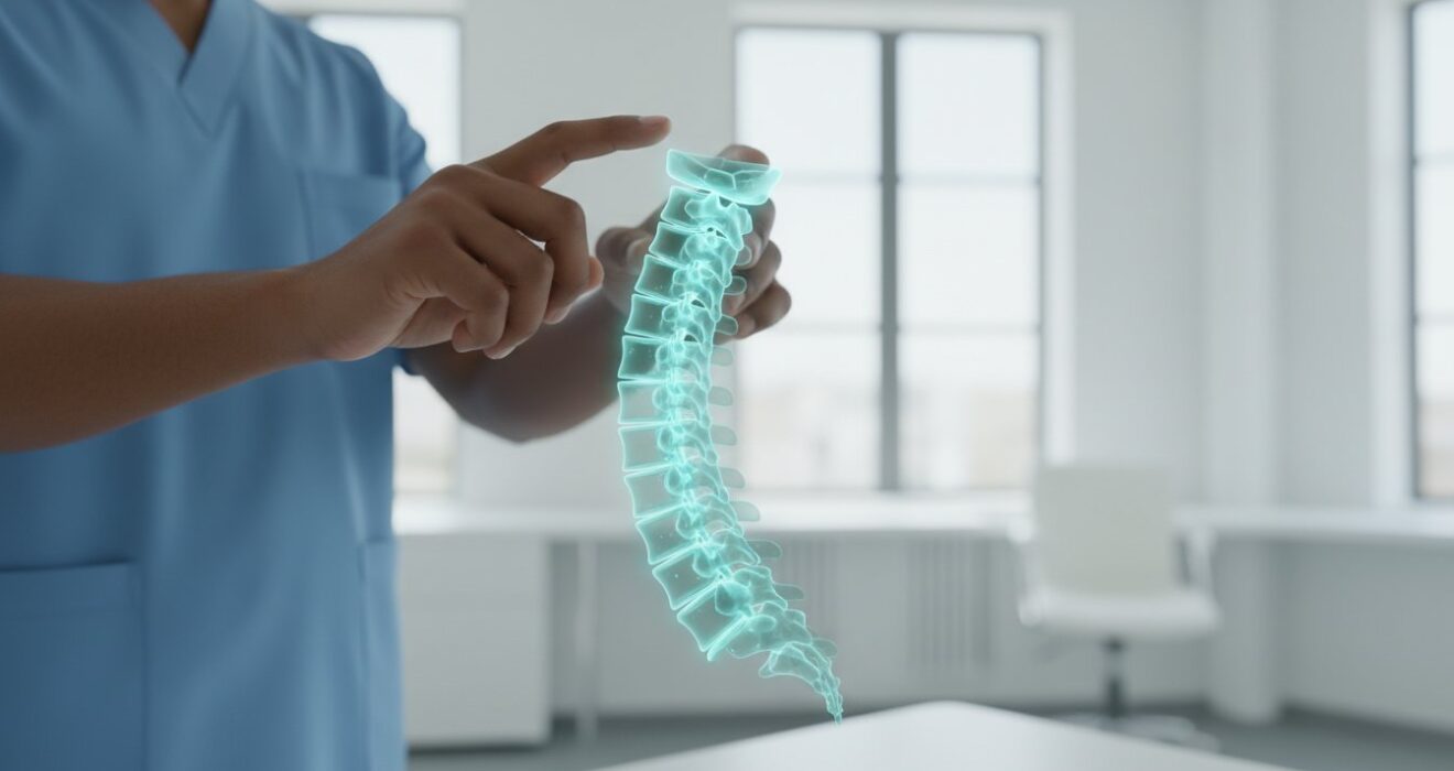
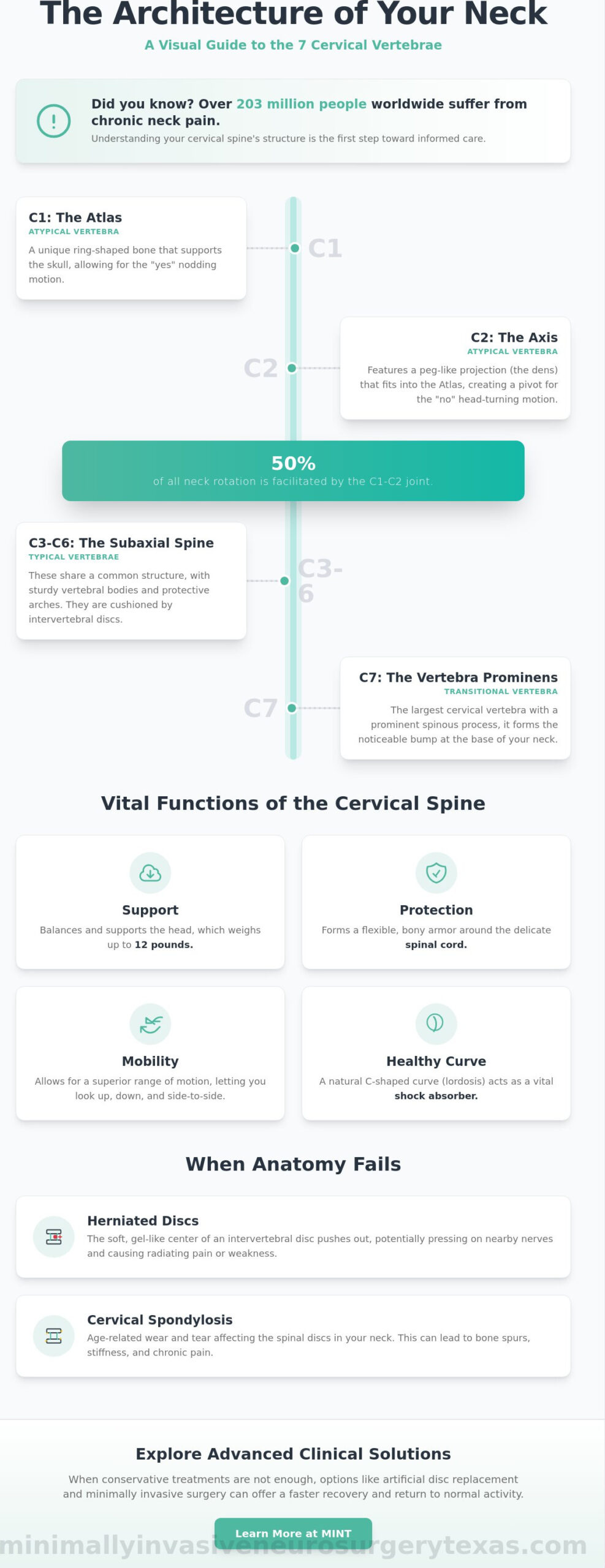
Did you know that over 203 million people worldwide currently suffer from chronic neck pain? This figure is projected to reach 269 million by 2050. When you experience stiffness or radiating discomfort, it’s natural to wonder about the structural integrity of your spine and exactly how many cervical vertebrae are there. Understanding this high-precision biological bridge is the first step toward reclaiming your daily quality of life.
You likely already feel that your neck is a complex masterpiece of engineering, yet it feels incredibly vulnerable when pain limits your movement. It’s common to feel overwhelmed by medical terminology or fear that your symptoms indicate a serious structural failure. We promise to help you discover the exact structure of your cervical spine and explain why these seven specific vertebrae are critical to your mobility and neurological health. This knowledge provides the clinical clarity you need to move from a state of concern to a state of informed confidence.
This guide explores the unique anatomy of the C1 through C7 vertebrae and details how they protect your spinal cord. You’ll learn about the vital roles of each segment and gain the knowledge necessary to discuss advanced options like artificial disc replacement or minimally invasive spine surgery with a neurosurgeon.
Key Takeaways
- Understand exactly how many cervical vertebrae are there and how their stacked structure facilitates the neck’s superior range of motion.
- Distinguish between the “atypical” C1 and C2 vertebrae and the “typical” bones of the mid-neck to pinpoint where your symptoms may originate.
- Learn how the cervical spine supports a head weighing up to 12 pounds while shielding the delicate spinal cord from potential injury.
- Discover how common conditions like cervical spondylosis or herniated discs can compromise these seven bones and disrupt your daily quality of life.
- Explore advanced clinical solutions, such as artificial disc replacement, that prioritize a fast recovery and a return to normal activity.
The Seven Cervical Vertebrae: An Overview of Your Neck’s Structure
The cervical spine is a marvel of biological engineering. It balances the heavy weight of the skull while providing the flexibility needed to scan your surroundings in an instant. When patients visit our practice seeking relief from chronic discomfort, they often ask about the basic anatomy of the region, specifically inquiring about how many cervical vertebrae are there and how they function. The answer is foundational to your health. There are exactly seven stacked bones in the neck, numbered C1 through C7. These vertebrae are significantly smaller than the larger, load-bearing lumbar vertebrae of the lower back. This compact size allows for the rapid, multi-directional head movement required for daily life.
The cervical spine is defined as the seven bones starting at the skull base and ending at the upper back. While they are the smallest vertebrae in the spinal column, they form its most mobile and flexible region. You can learn more about the specific characteristics of The Seven Cervical Vertebrae to understand how they differ from the rest of your back. This mobility makes them indispensable for your quality of life, but it also makes them susceptible to wear and tear over time.
The C1-C7 Numbering System Explained
Medical professionals use a standardized numbering system to communicate clearly about your spinal health. The “C” prefix stands for “cervical.” By labeling these bones from C1 at the top to C7 at the bottom, surgeons can pinpoint the exact origin of a patient’s symptoms. This precision is the first step in creating a personalized treatment plan for relief. The system ends where the neck meets the upper back. At this junction, C7 connects to T1, the first bone of the thoracic spine. Knowing if a pinched nerve is at the C5-C6 level versus the C6-C7 level determines the specific surgical approach required for your recovery.
Anatomical Landmarks of the Neck
You can actually feel the base of your cervical spine. If you run your hand down the back of your neck and tilt your head forward, you’ll notice a prominent bony bump. This is the C7 vertebra, often called the “vertebra prominens” because of its large spinous process. Beyond individual bones, the overall shape of the neck is equally important. A healthy cervical spine has a natural inward curve known as lordosis. This C-shaped curve acts as a vital shock absorber. It distributes the weight of your head evenly across the vertebrae and discs. When this curve is flattened or reversed, it often leads to chronic pain and structural instability.
C1 to C7: Understanding the Unique Roles of Each Vertebra
While you now know how many cervical vertebrae are there, it’s vital to recognize that these seven bones are not identical. They are divided into two distinct groups: the atypical vertebrae (C1 and C2) and the typical vertebrae (C3 through C6), with C7 acting as the final transition point. Each level performs a specialized task to ensure your head stays upright and your nervous system remains protected. Understanding these differences helps patients better describe their symptoms to a specialist.
The Atlas (C1) and Axis (C2): The Mobility Specialists
The top two levels of your spine are unique in their architecture. C1, known as the Atlas, is a ring-shaped bone that lacks a traditional vertebral body. It supports the globe of the skull, much like the mythological figure it’s named after. Directly beneath it sits C2, the Axis, which features a peg-like projection called the “dens.” This protrusion fits into the Atlas, creating a pivot joint that allows for significant flexibility. C1 and C2 facilitate 50% of all neck rotation. Because these bones sit so close to the brainstem, injuries at these levels are particularly critical for neurological function and require immediate, expert evaluation.
The Subaxial Spine: C3 through C7
The vertebrae from C3 through C6 share common structural features, such as a sturdy vertebral body and a protective arch. These bones are connected by intervertebral discs that act as cushions, allowing for smooth movement while preventing bone-on-bone contact. A deep understanding of cervical spine anatomy is essential for diagnosing why these discs might wear down or herniate over time.
Beyond structural support, these vertebrae perform several silent but vital functions:
- They house and protect the spinal cord within the vertebral canal.
- They feature small openings called transverse foramina that protect the vertebral arteries.
- These arteries are responsible for carrying oxygen-rich blood directly to the brain.
C7, the final segment, serves as a vital transition point between the highly mobile neck and the stable, rigid chest area. It’s built tougher than the bones above it to handle the shift in mechanical stress as it connects to the thoracic spine. When structural issues at any of these seven levels begin to cause radiating pain or numbness, it’s often a sign that the delicate balance of your anatomy has been disrupted. If you’re experiencing persistent symptoms, consulting a specialist about minimally invasive spine surgery can help you find a path back to pain-free movement.
Why Seven? The Vital Functions of the Cervical Vertebrae
While knowing how many cervical vertebrae are there provides a structural foundation, understanding their function reveals why specialized care is so critical. These seven bones perform a high-stakes balancing act every second of the day. They aren’t just a stack of supports; they’re a dynamic system designed to manage incredible physical demands while protecting your most vital biological pathways. From the moment you wake up, these vertebrae coordinate to provide a range of motion that no other part of the spine can match, including flexion, extension, and complex rotation.
The primary mechanical task of the cervical spine is supporting the weight of the human head. On average, an adult head weighs between 10 and 12 pounds. Because the neck is so flexible, these seven small bones must maintain stability even when the head is tilted at awkward angles, which can significantly increase the effective load on the spine. When this structural balance is lost, it often leads to the chronic pain and stiffness that bring patients to our practice seeking advanced clinical solutions.
Shielding the Central Nervous System
One of the most critical roles of the cervical vertebrae is the protection of the spinal cord. Each bone features a central opening called the vertebral foramen. When stacked together, these openings create a continuous, protective canal for the spinal cord as it exits the brainstem. The nerves at the cervical level are notably more vulnerable than those in the lower back because the canal is narrower and the nerves are responsible for essential functions in the arms and hands. If structural changes like disc herniation or bone spurs narrow this space, you might experience radiating arm pain, weakness, or “pins and needles” sensations. Our goal is always to restore this protective space and ensure your neurological health remains intact.
Blood Flow and the Vertebral Arteries
The cervical vertebrae possess a unique anatomical feature called the transverse foramina. These are small holes located on the sides of the vertebrae that serve as a dedicated passageway for the vertebral arteries. These arteries are vital. They carry oxygenated blood directly to the brain and cerebellum. This dual role makes the neck both a structural support and a vascular protector. If misalignments or significant bone growth occur, they can potentially impact cerebral circulation. This intersection of bone, nerve, and blood vessel is why we utilize high-precision diagnostic tools. It ensures that any intervention, whether it’s artificial disc replacement or another specialized procedure, preserves these critical life-support systems.
When Anatomy Fails: Common Conditions Affecting Cervical Vertebrae
Even a high-precision system like the cervical spine is susceptible to wear, trauma, and degenerative changes. While understanding how many cervical vertebrae are there provides a structural map, recognizing how these seven bones can fail is the first step toward relief. Most structural issues don’t occur randomly. They are concentrated at the C5-C6 and C6-C7 levels. These specific segments endure the highest mechanical stress because they sit at the base of the neck’s curve, bearing the weight of the head during every tilt and turn.
Common conditions often involve the breakdown of the intervertebral discs or the bones themselves. Cervical spondylosis is the natural wear and tear of the seven vertebrae that occurs as we age. This can lead to herniated discs, where the soft tissue between vertebrae C2 through C7 pinches an adjacent nerve. Additionally, stenosis involves the narrowing of the spinal canal, which can put direct pressure on the spinal cord and disrupt neurological signals. These conditions don’t just cause local stiffness; they can fundamentally alter your quality of life. Our patient’s guide to cervical vertebrae neck anatomy and modern care provides a detailed roadmap for understanding how these degenerative changes develop and what treatment options are available.
Foraminal Stenosis and Radiculopathy
When bone spurs develop on the vertebrae, they can narrow the small exits where spinal nerves branch out to the rest of the body. This is known as foraminal stenosis. It often leads to cervical radiculopathy, a condition where patients feel numbness, tingling, or weakness in their hands and fingers. A common point of confusion is that these symptoms often originate in the neck even if the neck itself doesn’t feel painful. This “sciatica-like” pain in the arm is a clear signal that one of the seven cervical levels is compromised and requires a professional evaluation.
The Connection to Neck Pain Diagnosis
Modern diagnostics allow us to move from uncertainty to a state of informed confidence. High-resolution imaging, such as an MRI or CT scan, maps your specific symptoms to one of the seven cervical vertebrae with absolute certainty. During a physical exam, we test the function of nerves at each level to confirm the imaging results. There’s a common misconception that all structural neck pain requires invasive, “open” surgery with long recovery times. In reality, many patients are excellent candidates for minimally invasive spine surgery. These advanced techniques focus on precision and tissue preservation, helping you achieve a faster return to normal activity without the trauma of traditional methods.
Advanced Care for Cervical Disorders in Plano and Lewisville
Your understanding of how many cervical vertebrae are there is the foundation of your diagnostic journey. However, anatomical knowledge is most valuable when it’s paired with a personalized clinical solution. For residents in Plano and Lewisville, chronic neck pain doesn’t have to be a permanent barrier to your daily quality of life. We transition from identifying structural issues to implementing high-tech treatments that prioritize your long-term mobility. Choosing a board-certified neurosurgeon ensures that your care is managed by a specialist who understands the intricate neurological pathways housed within these seven specific bones.
Local residents have direct access to sophisticated spinal navigation systems and robotic-assisted technology. These tools allow for a level of precision that was once impossible, ensuring that any intervention is tailored to your unique vertebral structure. Our commitment is to provide the relief of suffering through modern innovation, positioning our practice as a source of optimism for those dealing with degenerative conditions.
Minimally Invasive Solutions for the Cervical Spine
The modern standard for treating the neck has shifted toward motion preservation and tissue respect. One of the most effective options for eligible patients is neck disk replacement. This procedure replaces a damaged disc with an artificial one, maintaining the natural flexibility of the seven cervical vertebrae rather than fusing them together. By preserving motion, we reduce the stress on adjacent levels of the spine.
The shift toward minimally invasive neurosurgery has revolutionized patient outcomes. These techniques utilize small incisions and specialized instruments to reach the spine without significant disruption to the surrounding neck muscles. The benefits of this approach include:
- Reduced physical impact on the body during the procedure.
- Improved recovery timelines for a faster return to normal activity.
- Decreased postoperative discomfort compared to traditional open surgeries.
- Greater precision in addressing nerve decompression.
Consulting with MINT in North Texas
When you visit our Plano or Lewisville clinics, your evaluation begins with a comprehensive review of your health history and symptoms. Dr. Scott Kutz and the team at Minimally Invasive Neurosurgery of Texas (MINT) prioritize a patient-centered approach. We always explore non-surgical options first, utilizing surgery only when it’s the most effective path toward lasting relief. If an intervention is necessary, Dr. Kutz utilizes advanced technology like robotic navigation to treat vertebral issues with absolute certainty. This high-tech hallmark ensures that every movement is planned and executed with surgical excellence. Our goal is to move you from a state of concern to a state of informed confidence, helping you reclaim the active lifestyle you deserve.
Your Path to a Pain-Free Life and Restored Mobility
Understanding the complex engineering of your neck is the first step toward lasting relief. You now know exactly how many cervical vertebrae are there and why their structural integrity is vital for protecting your spinal cord and maintaining cerebral blood flow. When degenerative changes or injuries compromise these seven bones, the resulting pain can feel overwhelming. However, modern neurosurgical advancements offer a clear path forward through precision and innovation.
Led by board-certified neurosurgeon Dr. Scott Kutz, our practice specializes in motion-preserving artificial disc replacement and other advanced minimally invasive techniques. With convenient locations in Lewisville and Plano, TX, we prioritize your recovery and a swift return to normal activity. Don’t let chronic neck pain dictate your future. Schedule a Consultation with our Plano or Lewisville Spine Specialists Today to begin your journey toward informed confidence. We are here to provide the clinical excellence and compassionate care you need to move with comfort once again.
Frequently Asked Questions
What are the names of the 7 cervical vertebrae?
The seven cervical vertebrae are primarily identified by their alphanumeric labels, C1 through C7. The first two bones have specific names due to their unique shapes: C1 is called the Atlas and C2 is known as the Axis. The remaining bones, C3 through C7, are generally referred to by their numbers. This standardized system helps specialists communicate precisely about your spinal structure and the exact location of any symptoms.
Can you live with a broken cervical vertebra?
It’s possible to survive a broken cervical vertebra, but the outcome depends entirely on the stability of the fracture and whether the spinal cord is damaged. Stable fractures may only require bracing and observation. However, unstable injuries can lead to permanent neurological deficits or paralysis. If you suspect a neck injury, immediate immobilization and an evaluation by a neurosurgeon are essential to prevent further damage and ensure a safe recovery.
Which cervical vertebra is the most commonly injured?
The C2 (Axis) and C7 vertebrae are the most common sites for traumatic fractures. However, when discussing degenerative conditions like disc herniation or spondylosis, the C5-C6 and C6-C7 levels are the most frequently affected. These lower levels experience the highest amount of mechanical stress during daily movement. Understanding how many cervical vertebrae are there helps patients realize that the base of the neck bears the most weight and is therefore most prone to wear.
What is the difference between the Atlas and the Axis?
The Atlas (C1) is a ring-like bone that supports the weight of the skull, while the Axis (C2) features a bony projection called the “dens” that acts as a pivot point. While the Atlas allows for the “yes” nodding motion, the Axis is responsible for the “no” shaking motion. These two bones lack the traditional vertebral bodies found in the rest of the spine, making them specialized for superior flexibility and rotation.
Why is the C7 vertebra called the vertebra prominens?
C7 is called the vertebra prominens because it has a long spinous process that protrudes more than the other neck bones. This landmark is easily felt as a firm bump at the very base of your neck when you tilt your head forward. It serves as a vital clinical marker for surgeons and physicians to identify the transition point where the cervical spine meets the thoracic spine.
Do all mammals have seven cervical vertebrae?
Almost all mammals have exactly seven cervical vertebrae, regardless of the length of their necks. This includes species as diverse as humans, mice, and giraffes. The primary exceptions to this biological rule are manatees and two-toed sloths, which have six, and three-toed sloths, which have nine. For humans, these seven bones are the standard structural requirement for supporting the head and protecting the spinal cord.
How do I know if my neck pain is coming from a specific vertebra?
You can often identify the source of neck pain by observing where the symptoms radiate. For example, a problem at the C6 level typically causes pain or numbness in the thumb, while issues at C7 often affect the middle finger. Determining how many cervical vertebrae are there and mapping your symptoms to specific dermatomes is a process our team uses during a physical exam. We combine these clinical findings with MRI imaging to confirm the exact origin of your discomfort.