At Minimally Invasive Neurosurgery of Texas (MINT), board-certified neurosurgeon Dr. Scott Kutz provides specialized treatment for neural foraminal stenosis in Lewisville and Plano, offering advanced minimally invasive spine surgery to restore patient mobility. Dr. Kutz is a Fellow of the American Association of Neurological Surgeons and a Fellow of the American College of Surgeons.
What if the sharp, radiating pain in your arm or leg isn’t a permanent life sentence, but a treatable condition that doesn’t require invasive, open-back surgery? You’ve likely spent hours deciphering complex MRI reports or worrying that a surgical recommendation means a long, painful recovery. It’s common to feel frustrated when chronic pain limits your daily activities and mobility. Neural foraminal stenosis occurs when the small openings between the bones in your spine, known as the foramina, begin to narrow and compress the nerves passing through them. Dr. Scott Kutz uses precision-based, minimally invasive techniques to relieve this pressure and help you return to a normal activity level.
In this article, you’ll discover how specialized care at our Lewisville and Plano clinics provides a modern alternative to traditional spinal fusion. We will explain the common symptoms of nerve compression and how motion-preserving treatments can restore your quality of life.
Key Takeaways
- Learn why an MRI serves as the gold standard for diagnosing neural foraminal stenosis and how Dr. Scott Kutz uses these images to identify nerve root encroachment.
- Identify the hallmark signs of radiculopathy and understand why symptoms like radiating pain typically occur on only one side of the body.
- Review conservative care options such as anti-inflammatory medications and physical therapy that often provide relief before surgery is necessary.
- Explore how Dr. Scott Kutz, board-certified neurosurgeon, Fellow of the American Association of Neurological Surgeons, and Fellow of the American College of Surgeons, utilizes specialized minimally invasive spine surgery at MINT to restore mobility.
- Discover the benefits of seeking treatment at our state-of-the-art facilities in Lewisville and Plano to begin your journey back to a normal activity level.
Minimally Invasive Neurosurgery of Texas (MINT) and Dr. Scott Kutz provide specialized care for neural foraminal stenosis in Lewisville and Plano, TX. This specific diagnosis is often a source of confusion for patients reading their MRI results for the first time. Neural foraminal stenosis involves the narrowing of the small openings called foramina where nerve roots exit the spinal canal. While Spinal stenosis is a broad medical term for any narrowing within the spine, foraminal stenosis specifically targets the exit portals on the sides of the vertebrae.
It is vital to distinguish this from central canal stenosis. In central canal stenosis, the narrowing occurs in the main channel that houses the spinal cord itself. Conversely, foraminal stenosis affects the individual nerve roots after they have already branched off from the spinal cord. Patients often experience different symptoms depending on which type of compression they have. Dr. Scott Kutz, a board-certified neurosurgeon and Fellow of the American Association of Neurological Surgeons, uses advanced imaging to pinpoint exactly where the pressure occurs. This precision allows the team at MINT to apply minimally invasive spine surgery techniques that target the specific point of compression without disturbing healthy surrounding tissue.
The Anatomy of the Foramen
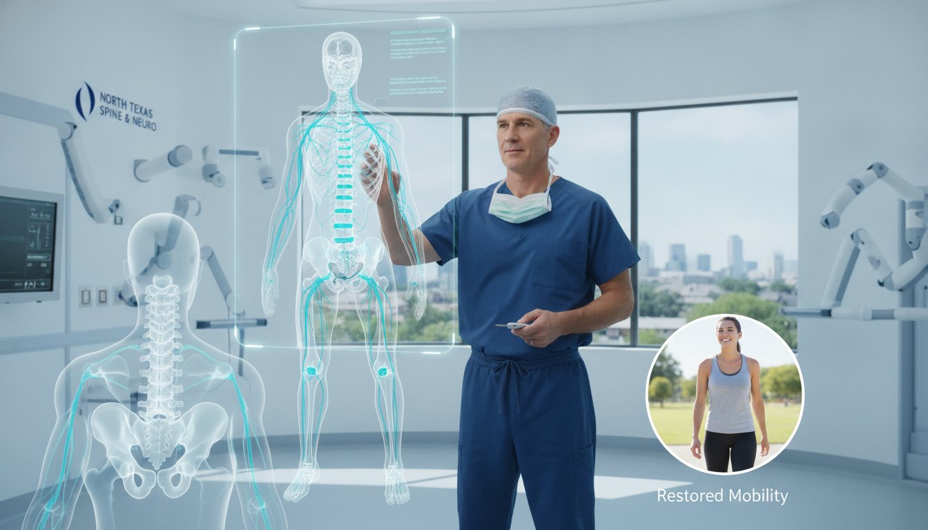
The neural foramen serves as a protective passageway. Its primary role is to shield the delicate nerve roots as they travel from the spinal cord to your limbs. You can visualize each foramen as a doorway between two stacked vertebrae. The boundaries of this doorway consist of the vertebral bones, the facet joints, and the intervertebral discs. When these structures change due to aging or injury, the doorway becomes smaller. This narrowing is what leads to nerve impingement. As the space shrinks, the nerve loses its protective cushion, resulting in the “pinched nerve” sensation that drives many patients to seek a specialist in Lewisville or Plano.
Why Location Matters: Cervical vs. Lumbar Stenosis
The location of the narrowing dictates where you will feel pain. The lower back, known as the lumbar spine, remains the most frequent site for this condition. This area supports the majority of your body weight, making it susceptible to the degenerative changes that cause narrowing. Lumbar compression typically causes pain to radiate into the legs or feet. In contrast, cervical stenosis occurs in the neck and sends pain, tingling, or weakness down into the arms and hands. While thoracic stenosis in the mid-back is possible, it occurs much less frequently because the rib cage keeps those vertebrae relatively stable. Understanding these regional differences helps Dr. Scott Kutz develop a tailored treatment plan at Minimally Invasive Neurosurgery of Texas to restore your mobility.
Symptoms and Causes of Foraminal Narrowing
Many patients who visit our Lewisville or Plano offices describe a sharp, electric sensation that travels down a single arm or leg. This condition, known as radiculopathy, serves as the hallmark symptom of neural foraminal stenosis. Because the spine contains two exit portals at every level, narrowing usually occurs on just one side. This explains why your symptoms are likely unilateral, meaning they affect only the right or left side of your body rather than both simultaneously.
Dr. Scott Kutz, a board-certified neurosurgeon, Fellow of the American Association of Neurological Surgeons, and Fellow of the American College of Surgeons, often observes that patients initially mistake these signals for simple muscle strain. However, a muscle injury typically heals with rest, while nerve compression behaves differently. Certain movements, such as standing for long periods or walking, often increase the pressure within the foramen. You might find that leaning forward or sitting provides temporary relief because these positions slightly open the spinal passageways and reduce the immediate pinch on the nerve root.
Common Symptoms to Watch For
The early stages of nerve root encroachment often manifest as sensory changes that disrupt your daily routine. You should monitor for these specific warning signs:
- Burning sensations that feel like a localized heat or a sharp zap along the nerve path.
- Tingling or a “pins and needles” feeling that persists in the hands, feet, or fingers.
- Muscle weakness that makes it difficult to grip objects or lift your foot while walking.
- Loss of coordination or a heavy feeling in the limbs that leads to frequent tripping.
While many cases of Foraminal Stenosis progress slowly, some symptoms require immediate intervention. If you experience a sudden loss of bowel or bladder control, you must seek emergency medical care immediately as this indicates severe neurological compromise.
The Primary Causes of Stenosis
Several structural changes lead to the narrowing of the neural foramen. Degenerative disc disease is a leading contributor; as discs lose height and water content, the space between the vertebrae collapses and shrinks the exit portal. In response to this instability, the body often creates bone spurs, or osteophytes. These hard growths can protrude directly into the foramen space and compress the nerve.
Other factors include thickened ligaments, which lose their elasticity and bulge into the spinal canal, and spondylolisthesis, a condition where one vertebra slips forward over another. Dr. Scott Kutz utilizes state-of-the-art robotic navigation at Minimally Invasive Neurosurgery of Texas to identify these specific structural triggers with absolute precision. If you’re concerned about persistent radiating pain, you can schedule a consultation to discuss your diagnostic imaging and explore your options for relief.
Diagnosing Severity: Mild to Severe Foraminal Stenosis
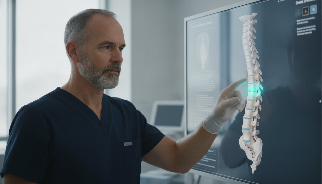
Magnetic Resonance Imaging (MRI) serves as the gold standard for visualizing nerve root encroachment in patients suffering from neural foraminal stenosis. This imaging technology provides high-resolution views of soft tissues, allowing Dr. Scott Kutz, a board-certified neurosurgeon, Fellow of the American Association of Neurological Surgeons, and Fellow of the American College of Surgeons, to see exactly how much space remains for the nerve. However, Dr. Kutz emphasizes that an image only tells half the story. He prioritizes patient-first communication at Minimally Invasive Neurosurgery of Texas to ensure you understand that your symptoms must correlate with what appears on the screen.
It’s quite common for a patient to feel alarmed by the word “severe” on a radiology report. Interestingly, some patients with severe narrowing experience fewer daily symptoms than those with a “mild” classification. This occurs because the human body often adapts to slow structural changes over time. A thorough physical exam remains the most critical step in the diagnostic journey. Dr. Kutz evaluates your muscle strength, reflexes, and pain patterns to confirm if the narrowing seen on the MRI is the actual source of your discomfort. This clinical correlation ensures that we target the correct area during any future minimally invasive spine surgery.
Understanding Your MRI Report
When you review your results at our Lewisville or Plano clinic, you will likely see the severity graded into three categories. Mild narrowing indicates that the foramen is slightly smaller, but there is no contact with the nerve root. Moderate narrowing means the space is restricted enough that it may touch the nerve root, but it does not push it out of its natural path. Severe narrowing represents significant compression, where the surrounding bone or disc material clearly displaces the nerve root from its normal position.
Diagnostic Tools Beyond Imaging
While an MRI provides the visual evidence, other tools help confirm how the nerve is actually functioning. Dr. Scott Kutz may recommend these additional tests:
- Electromyography (EMG): This test measures the electrical activity of muscles and the nerves that control them to identify the extent of nerve damage.
- Diagnostic Nerve Blocks: An injection of anesthetic near the suspected nerve helps confirm the exact source of pain. If the pain temporarily vanishes, the diagnosis is confirmed.
- X-rays: These images help Plano patients check for spinal instability or spondylolisthesis, which is when one vertebra slips over another.
Treatment Pathways: From Conservative Care to Minimally Invasive Surgery
Dr. Scott Kutz generally recommends starting with conservative management for neural foraminal stenosis before considering surgical intervention. This initial phase often includes targeted physical therapy to strengthen the muscles supporting the spine and nonsteroidal anti-inflammatory drugs (NSAIDs) to manage daily discomfort. These methods aim to reduce the mechanical stress on the narrowed foramen and improve your overall functional mobility without the need for an operating room.
If oral medications and therapy don’t provide sufficient relief, epidural steroid injections serve as a powerful secondary option. These injections deliver concentrated anti-inflammatory medication directly into the epidural space near the compressed nerve root. While an injection doesn’t physically widen the narrowed opening, it significantly reduces nerve swelling and inflammation. This often creates a window of relief that allows patients to progress further in their physical rehabilitation. When these conservative efforts fail to resolve radiating pain after several weeks, Dr. Kutz explores specialized Minimally Invasive Spine Techniques to address the underlying structural issue.
The Minimally Invasive Advantage
The primary benefit of a minimally invasive approach lies in its precision. Dr. Scott Kutz, a board-certified neurosurgeon, Fellow of the American Association of Neurological Surgeons, and Fellow of the American College of Surgeons, utilizes muscle-sparing techniques that avoid the large, traumatic incisions required in traditional open back surgery. Instead of cutting through healthy tissue, he uses specialized dilators to gently separate muscle fibers. This methodology results in significantly less blood loss and a lower risk of postoperative infection. Because the procedure minimizes trauma to the surrounding area, most MINT patients in Lewisville and Plano return home the same day or shortly after their procedure.
Advanced Surgical Options
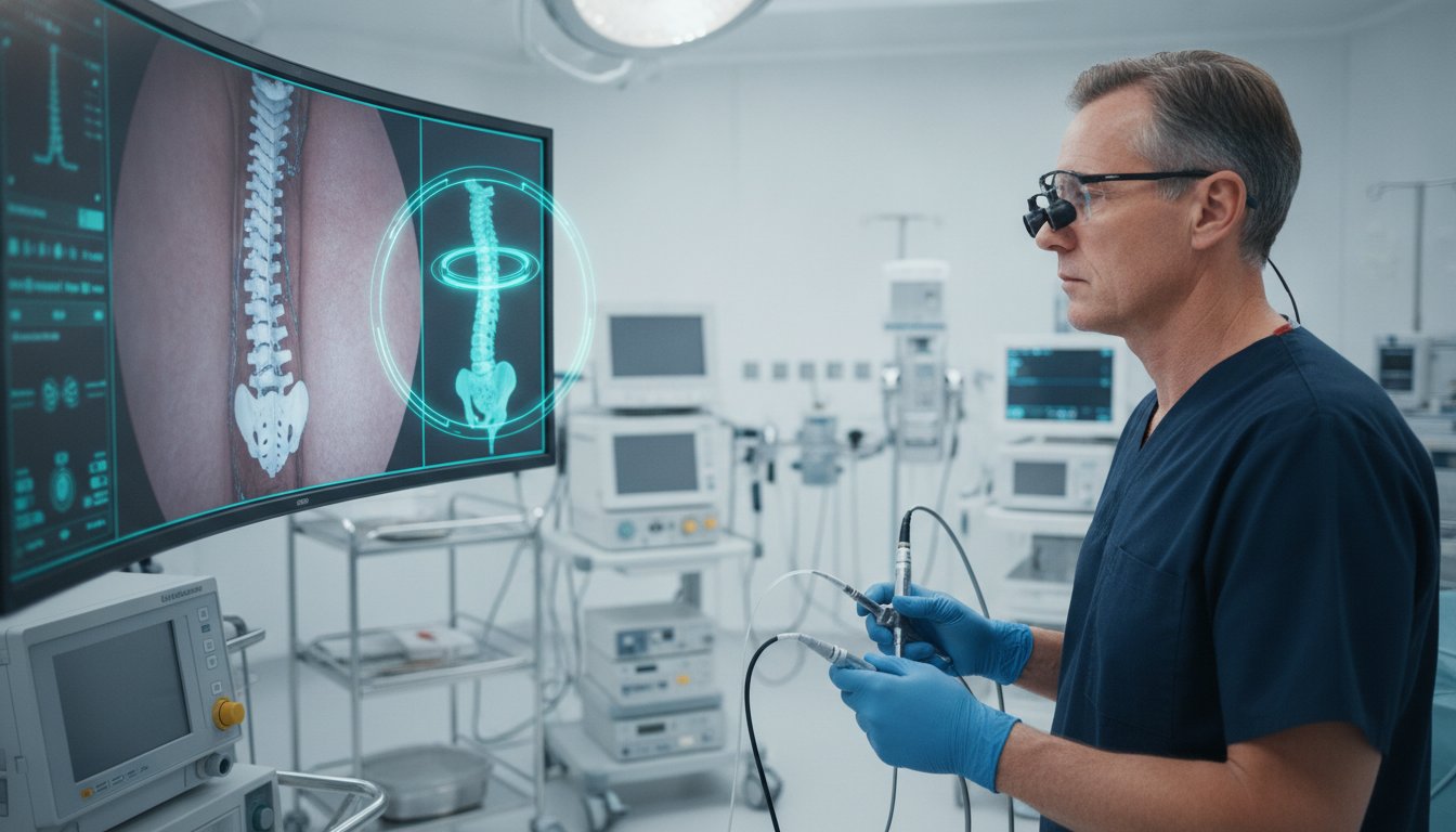
At Minimally Invasive Neurosurgery of Texas, we utilize cutting-edge technology like the Globus Excelsius robotic navigation system and Augmedics AR to enhance surgical accuracy. For patients requiring decompression, Endoscopic Spine Surgery allows Dr. Kutz to remove bone spurs or herniated disc material through a tiny tube with ultra-precise visualization. In cases involving the cervical spine, Artificial Disc Replacement offers a superior alternative to traditional fusion. While fusion locks two vertebrae together and limits movement, a replacement disc preserves your natural range of motion and protects the health of adjacent spinal levels. If you’re ready to move beyond temporary fixes, you can schedule a consultation at our Lewisville or Plano office to discuss these advanced motion-preserving options.
Minimally Invasive Solutions at MINT: The Texas Advantage
Dr. Scott Kutz, board-certified neurosurgeon, Fellow of the American Association of Neurological Surgeons, and Fellow of the American College of Surgeons, provides a level of precision that traditional spine centers often struggle to match. At Minimally Invasive Neurosurgery of Texas (MINT), we focus on treating neural foraminal stenosis with a blend of clinical expertise and the latest surgical innovations. Our practice operates as a specialized boutique center. We prioritize personal attention over the high volume often seen in massive, impersonal hospital networks.
A key part of our success involves the use of the Globus Excelsius robotic navigation system. This technology allows Dr. Kutz to plan your procedure with sub-millimeter accuracy before the first incision is ever made. During the surgery, the Augmedics AR augmented reality headset acts as a high-tech GPS for the surgeon. It overlays your specific spinal anatomy directly onto the surgeon’s field of view. This ensures that every movement is exact, protecting your delicate nerve roots and speeding up your return to normal activity.
Personalized Care in Lewisville and Plano
Our patient-centered philosophy means we listen to your goals and concerns before suggesting a treatment path. Following our 2026 headquarters relocation to Lewisville, we’ve expanded our ability to serve the North Texas community with state-of-the-art facilities. While our primary clinics are in Lewisville and Plano, we also care for patients from Dallas, Fort Worth, and McKinney. We encourage you to view our Patient Testimonials to see how our motion-preserving techniques have helped others regain their independence and quality of life.
Preparing for Your Consultation
Coming prepared to your appointment helps us move quickly toward a solution. Please bring your most recent MRI or CT scans on a disc so Dr. Kutz can review the raw images rather than just the written report. You should feel empowered to ask specific questions during your visit, such as:
- Is my condition a candidate for a non-fusion procedure?
- What is the expected recovery timeline for endoscopic surgery?
- How will this procedure improve my specific radiating limb pain?
- Are there lifestyle changes I can make to support my recovery?
If you’re tired of living with the limitations of nerve compression, it’s time to seek a definitive diagnosis from a specialist who understands your pain. Contact Minimally Invasive Neurosurgery of Texas today to schedule your evaluation at our Lewisville or Plano office and start your journey back to a pain-free life.
Take the Next Step Toward Lasting Nerve Relief
Living with persistent nerve pain often feels overwhelming, but modern medicine offers a clear path toward relief. You now understand that neural foraminal stenosis involves specific structural narrowing that requires a precision-based approach. Dr. Scott Kutz, a board-certified neurosurgeon, Fellow of the American Association of Neurological Surgeons, and Fellow of the American College of Surgeons, utilizes advanced robotic navigation and augmented reality to provide ultra-precise decompression. This high-tech care at our Lewisville and Plano facilities prioritizes your long-term mobility and a return to normal activity.
By choosing a specialized center like MINT, you avoid the impersonal nature of large hospital systems. We focus on motion-preserving alternatives to traditional fusion, helping you maintain your natural spinal function. It doesn’t have to be a struggle to find local, expert care in North Texas. Our team stands ready to help you move past chronic pain and reclaim your quality of life.
Schedule Your Consultation with Dr. Scott Kutz Today and take the first step toward lasting relief. You deserve a recovery plan built around your unique needs and goals.
Frequently Asked Questions about Neural Foraminal Stenosis
Is neural foraminal stenosis a serious condition?
This condition becomes serious when it leads to progressive neurological deficits such as muscle weakness or a loss of coordination. While mild cases are often manageable with conservative care, severe compression can cause permanent nerve damage. Dr. Scott Kutz, a board-certified neurosurgeon and Fellow of the American Association of Neurological Surgeons, evaluates each case to prevent long-term disability through early intervention.
Can foraminal stenosis be cured without surgery?
You can successfully manage the symptoms of neural foraminal stenosis without surgery, but the physical narrowing of the bone does not reverse on its own. Conservative treatments like anti-inflammatory medications and steroid injections focus on reducing nerve swelling. These methods help many patients in Lewisville and Plano return to their daily routines without requiring an operation.
What happens if neural foraminal stenosis is left untreated?
Leaving nerve compression untreated can lead to chronic, debilitating pain and permanent loss of function. Over time, the constant pressure may cause muscle atrophy, which is the wasting away of muscle tissue due to lack of nerve stimulation. Patients may also experience a permanent loss of sensation or “numbness” in the affected arm or leg.
How long is the recovery after minimally invasive surgery for stenosis?
Most patients at our Lewisville and Plano clinics return home the same day as their procedure. You can typically resume light daily activities within one to two weeks. Full recovery and a return to more strenuous physical tasks usually occur within six to twelve weeks as the internal tissues heal completely.
Does foraminal stenosis always cause pain?
Narrowing of the spinal openings does not always result in physical pain. Some individuals show “moderate” or even “severe” narrowing on an MRI but remain asymptomatic because the nerve root still has enough clearance to avoid irritation. Dr. Scott Kutz only recommends treatment when your clinical symptoms directly correlate with your diagnostic imaging.
What is the success rate of endoscopic spine surgery for stenosis?
Recent data from 2026 indicates that 80% to 95% of patients experience significant pain relief and improved function following minimally invasive spine surgery. Dr. Scott Kutz utilizes advanced robotic navigation and endoscopic tools to achieve these high success rates. This precision-based approach ensures the best possible outcome for patients throughout North Texas.
Can physical therapy reverse foraminal narrowing?
Physical therapy cannot physically widen the bone openings or shrink bone spurs associated with neural foraminal stenosis. It is, however, highly effective at strengthening the supporting muscles and improving spinal alignment. This reduces the mechanical pressure on the nerves and often allows patients to avoid surgery entirely by stabilizing the spine.
Will I need a spinal fusion for neural foraminal stenosis?
You do not always need a spinal fusion to treat this condition. Dr. Scott Kutz, board-certified neurosurgeon and Fellow of the American College of Surgeons, prefers motion-preserving alternatives like artificial disc replacement or simple decompression. These advanced techniques at MINT relieve the nerve pressure while maintaining your natural flexibility and preventing the stiffness of a fusion.