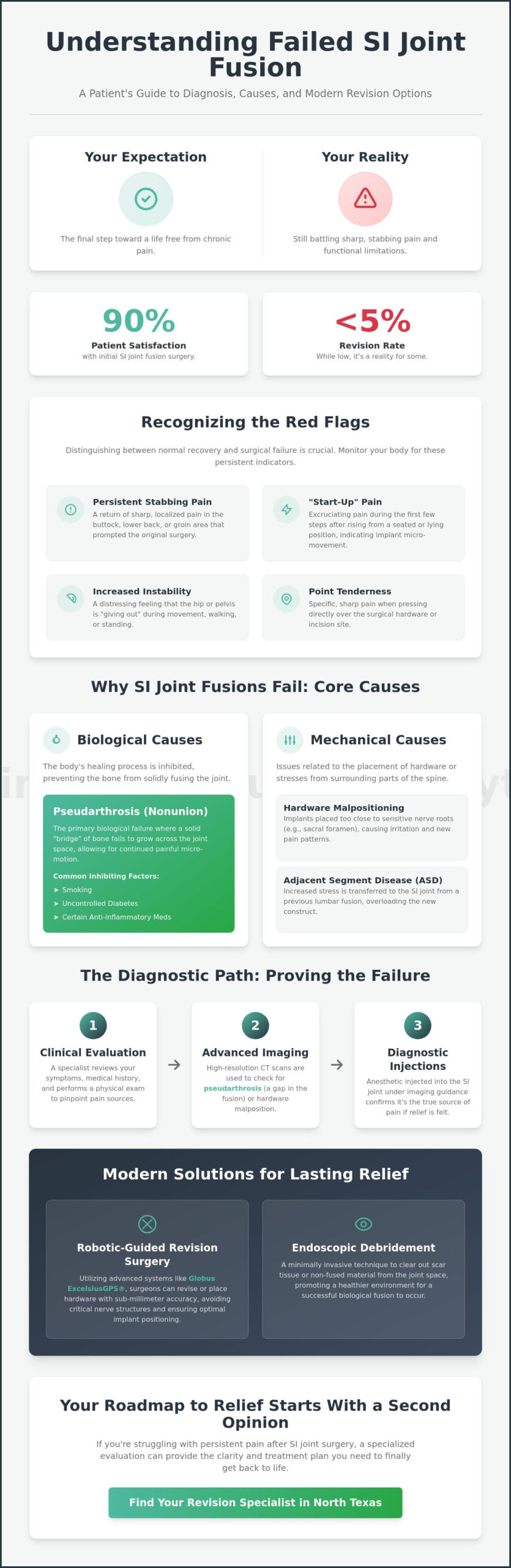
A “failed” SI fusion is often a diagnostic or mechanical error, not a permanent sentence of chronic pain. You likely expected your surgery to be the final step toward recovery, yet you’re still battling that sharp, stabbing sensation in your buttock and lower back. It’s exhausting to hear that your imaging looks “normal” when you still can’t sit for more than ten minutes or walk comfortably, despite your commitment to the healing process. While revision rates for these procedures remain below 5% in 2026 clinical trials, that statistic doesn’t change the reality of your daily struggle.
We promise to help you identify the specific symptoms of failed si joint fusion and provide a clear roadmap to the relief you deserve. By understanding the criteria outlined in the January 2026 Carelon clinical guidelines, you can determine if your pain stems from implant malposition, nonunion, or an incorrect initial diagnosis. This article explores how modern MISS techniques and advanced robotic navigation can correct these issues, ensuring you have the information needed to find a specialist and truly get back to life.
Key Takeaways
- Learn to distinguish between normal post-operative recovery and the persistent symptoms of failed si joint fusion, such as stabbing buttock pain or hip instability.
- Discover the biological and mechanical reasons why fusions fail, including pseudarthrosis and hardware malpositioning that irritates nearby nerve roots.
- Understand the diagnostic path to proving a failure, utilizing high-resolution CT scans and diagnostic injections to pinpoint the exact source of your discomfort.
- Explore innovative revision options like robotic-guided surgery with Globus Excelsius and endoscopic debridement designed for faster recovery and precision.
- Find out how a specialized second opinion from a board-certified neurosurgeon in North Texas can provide the roadmap you need to get back to life.
Recognizing the Red Flags: Symptoms of Failed SI Joint Fusion
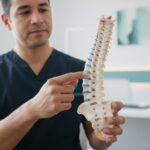
The primary goal of a fusion procedure is to stabilize the joint and eliminate the debilitating effects of Sacroiliac joint dysfunction. When the surgery is successful, the joint becomes a solid bridge of bone, ending the painful friction between the sacrum and the ilium. However, when the procedure doesn’t achieve this intended result, the body sends clear signals. The most common symptoms of failed si joint fusion involve a return of the sharp, localized pain that prompted the surgery in the first place, or occasionally, the development of entirely new pain patterns.
Patients living with a failed fusion often report specific physical sensations that differ from standard post-operative soreness. You should monitor your body for these indicators:
- Persistent or “new” stabbing pain localized in the buttock or groin area.
- A distressing feeling of increased instability, often described as the hip “giving out” during a stride.
- Sharp pain that radiates down the leg, which many patients mistake for sciatica or a lumbar disc issue.
- Point-specific tenderness directly over the surgical hardware or the original incision site.
Normal Recovery Pain vs. Surgical Failure
Distinguishing between healing and failure requires patience and clinical insight. A standard biological fusion typically follows a timeline of 3 to 6 months for the bone to grow across the joint space completely. During the first few weeks, some discomfort is expected as your body reacts to the hardware. A red flag appears when this discomfort does not diminish or if it suddenly intensifies after a period of improvement. Failed back surgery syndrome, as it relates to the SI joint, is the clinical term used when a patient does not achieve the expected pain relief or functional improvement following a sacroiliac fusion surgery.
The “Start-Up” Pain Phenomenon
One of the most diagnostic symptoms of failed si joint fusion is “start-up” pain. This is a specific type of mechanical distress that occurs when you rise from a chair or get out of bed. If the implants have loosened or failed to integrate with the bone, the sudden shift in weight-bearing forces causes the hardware to micro-move. This movement irritates the surrounding nerve-rich tissues, making the first few steps of any activity excruciating. While 88% to 90% of patients are satisfied with their initial surgery, those experiencing mechanical loosening often find that their symptoms worsen during any weight-bearing activity, signaling that the joint is not yet truly stable.
Why SI Joint Fusions Fail: Biological and Mechanical Causes
Understanding why a procedure fails is the first step toward a successful revision. While success rates for sacroiliac fusion are historically high, reaching 80% to 90% in recent clinical studies, a small percentage of patients don’t achieve the expected relief. These failures typically fall into two categories: biological and mechanical. When the body doesn’t cooperate with the hardware, or the hardware itself is poorly positioned, the symptoms of failed si joint fusion begin to surface, often mirroring the original pain that led you to surgery. To better understand what a successful procedure looks like and how it differs from a failed one, reviewing a comprehensive overview of si joint fusion surgery can help you identify where your recovery may have gone wrong.
A significant factor we see in our North Texas clinics is Adjacent Segment Disease. Patients who have previously undergone lumbar fusions often experience a transfer of stress to the sacroiliac joint. If an SI fusion is performed without addressing this compensatory stress, or if the joint fails to stabilize, the mechanical load continues to cause pain. Additionally, hardware malpositioning can occur when implants are placed too close to the sacral foramen, irritating sensitive nerve roots. If you suspect your recovery has stalled, a specialized second opinion can help clarify whether your pain is mechanical or biological in nature.
The Role of Pseudarthrosis
Fusion is a biological process, not just a mechanical one. Pseudarthrosis occurs when the bone fails to grow across the joint space, leaving the joint mobile despite the presence of implants. Research into the causes of SI joint pain after fusion indicates that lifestyle factors play a massive role here. Smoking, uncontrolled diabetes, and certain anti-inflammatory medications can inhibit osteoblast activity. When this bridge of bone doesn’t form, micro-motion continues. This tiny, repetitive movement triggers pain receptors constantly, preventing you from getting back to life.
Mechanical Loosening and Hardware Failure
Mechanical failure involves the implants themselves. Even with world-class titanium hardware, integration isn’t guaranteed. Stress shielding is a common phenomenon where the metal implant carries too much of the physical load, causing the surrounding bone to lose density because it isn’t being “worked.” This leads to loosening of the surgical screws. In rare cases, the hardware may even fracture under the intense pressure of the pelvic girdle. While radiographic fusion rates are 96% at 12 months for most patients, those with mechanical loosening will feel a distinct grinding or shifting sensation during weight-bearing activities. If you’re new to understanding spinal procedures, learning what is back fusion surgery and how modern techniques achieve stability can help you ask better questions during your revision consultation. Identifying these symptoms of failed si joint fusion early allows for more effective intervention using advanced navigation technology.
The Diagnostic Path: Proving the Failure
Confirming a surgical failure requires a sophisticated diagnostic protocol that goes far beyond a standard office visit. Many patients arrive at our clinic feeling gaslit by previous providers who dismissed their pain because post-operative imaging appeared “normal.” Proving a failure involves a multi-step verification process to ensure the symptoms of failed si joint fusion are correctly attributed to the joint itself rather than a neighboring lumbar issue. This process is essential for building a predictable path toward a successful revision and helping you finally get back to life.
Our diagnostic journey typically begins with provocative physical testing. During an examination, a neurosurgeon performs specific maneuvers like the FABER (Flexion, Abduction, and External Rotation) test or Gaenslen’s test. These movements are designed to stress the sacroiliac joint specifically. If these tests reproduce your typical stabbing pain, it provides the first clinical evidence that the fusion has not achieved the necessary stability. We then pair these findings with dynamic X-rays, which allow us to observe the hardware while your body is in motion, looking for any signs of screw migration or loosening that might be invisible on static films.
Why Standard MRIs Often Miss Failed Fusions
Standard MRIs are frequently the first tool ordered by general practitioners, yet they are often the least helpful in this specific context. The presence of metal titanium implants creates “artifacting,” a phenomenon where the hardware distorts the magnetic field and blurs the surrounding tissue. This often leads to a radiology report that misses the subtle symptoms of a failed SI joint fusion, such as a lack of bone bridging. We consider high-resolution CT scans the gold standard because they provide the clarity needed to see if a biological bridge has actually formed. A surgeon-read CT scan is vital for precision, as it allows us to inspect the interface between the bone and the implant with microscopic detail.
The Diagnostic Injection Test
The most definitive tool in our diagnostic arsenal is the controlled injection. We use fluoroscopic guidance to place a numbing agent directly into the joint space or around the hardware. To confirm the SI joint as the pain source, we look for the “80% relief rule.” If you experience 80% or more reduction in your typical pain for the duration of the anesthetic, it confirms that the joint is the primary culprit. This test is crucial because it differentiates SI joint distress from lumbar disc herniations or hip pathology. Diagnostic blocks are the most reliable way to map surgical success. When these injections provide temporary but significant relief, they validate that the symptoms of failed si joint fusion are treatable through a specialized revision procedure.
Revision Options: Minimally Invasive Solutions at MINT
If you’re struggling with the symptoms of failed si joint fusion, you need a solution that addresses the physical reality of your condition. Many alternative clinics suggest non-surgical “prolotherapy” as a catch-all fix, but these injections cannot tighten a loose screw or correct malpositioned hardware. When a mechanical failure occurs, a mechanical correction is necessary. At Minimally Invasive Neurosurgery of Texas, we specialize in revision procedures that utilize advanced technology to correct previous errors with microscopic precision, allowing you to finally get back to life.
Revision surgery doesn’t always mean a total removal of existing hardware. In many cases, we can perform a supplemental fusion. This involves adding stability to the joint through a different trajectory or using a different implant system without the trauma of extracting well-integrated screws. By utilizing 3D-printed implants, which showed a 72% bridging fusion rate at just 6 months in January 2026 studies, we can encourage biological growth where the first surgery failed. This targeted approach is the cornerstone of Minimally Invasive Neurosurgery: The Modern Standard for Spine Care in 2026, ensuring that revision is a path to resolution rather than just another operation.
The Advantage of Robotic Navigation
Precision is the most critical factor in revision surgery. Robotic-guided revision using the Globus Excelsius system allows us to map your unique pelvic anatomy in three dimensions before we ever make an incision. This technology prevents further nerve irritation by ensuring that new hardware avoids the sacral foramen entirely. Clinical data from 2026 indicates that radiographic fusion rates reach 100% by 18 months when robotic navigation is employed. Because this is a MISS procedure, you’ll experience significantly less blood loss and muscle trauma compared to traditional open revisions, leading to a faster and more predictable recovery.
Non-Surgical Management for Borderline Cases
Not every patient experiencing the symptoms of failed si joint fusion requires immediate surgery. For borderline cases where the hardware is stable but the patient still feels nerve-related distress, radiofrequency ablation (RFA) can be an effective bridge. This procedure uses thermal energy to “turn off” the pain signals from the small sensory nerves around the SI joint. We also implement specialized physical therapy protocols focused on gait correction. After a failed fusion, many patients develop compensatory walking patterns that strain the lower back. Correcting these mechanics, sometimes paired with biological stimulants to encourage bone growth, can occasionally resolve pain without further invasive steps.
If your previous surgery hasn’t delivered the results you were promised, it’s time for a more precise perspective. Schedule a revision consultation today to explore how our robotic-guided techniques can restore your mobility.
Finding Relief in North Texas: Choosing a Revision Specialist
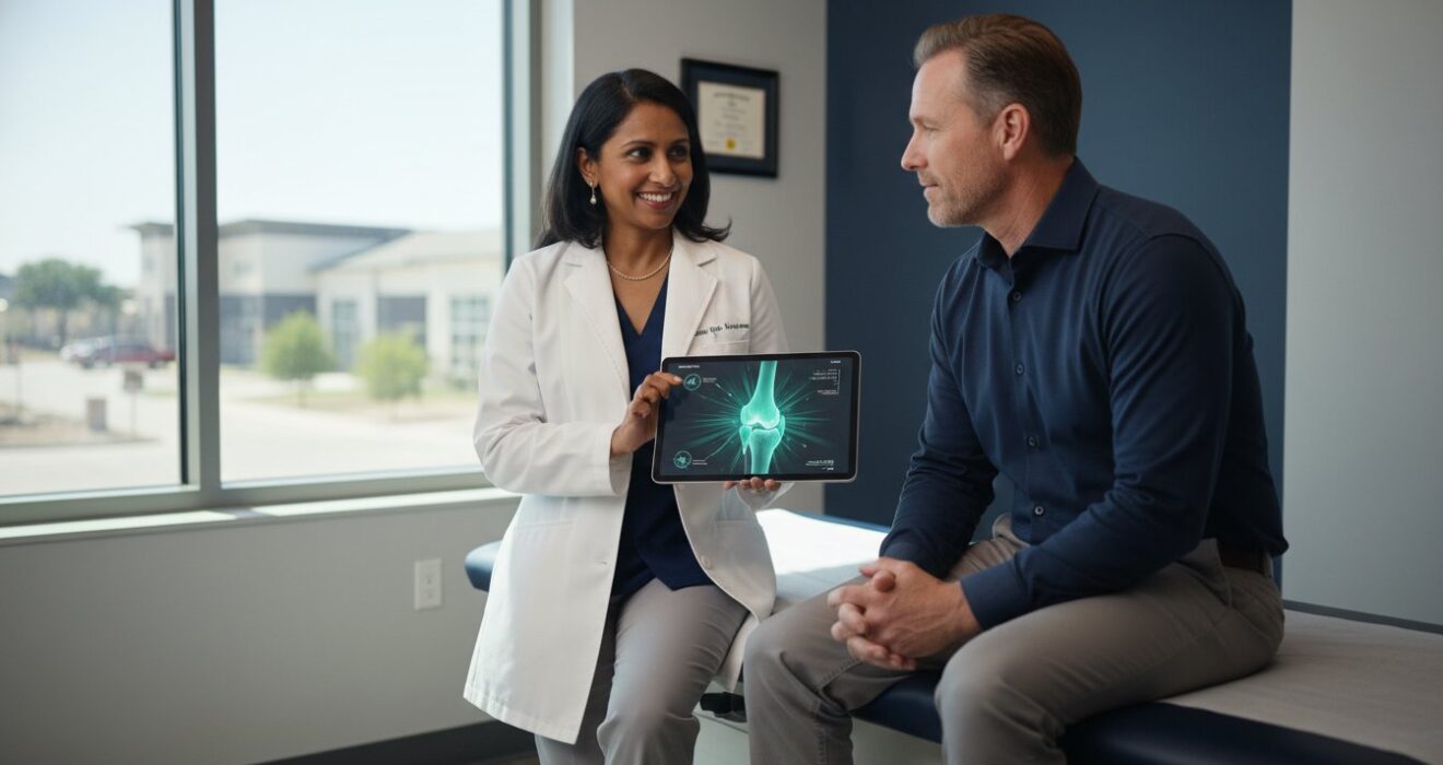
When your initial surgery doesn’t provide the relief you were promised, the path forward requires more than just another procedure. It demands a specialized level of board-certified neurosurgical expertise. Dr. Scott Kutz and the team at Minimally Invasive Neurosurgery of Texas understand that a second opinion isn’t just a formality; it’s a vital diagnostic step for any patient struggling with the symptoms of failed si joint fusion. By re-evaluating your case through the lens of a MISS specialist, we can often identify subtle mechanical or biological issues that were previously overlooked.
Our approach is rooted in the belief that every patient deserves a personalized roadmap to recovery. While revision surgery for failed SI joint fusion remains uncommon, with rates below 5% in large 2026 clinical trials, those who do require it need a surgeon who is a master of both anatomy and technology. We prioritize your health by combining a warm, patient-centered environment with the high-tech precision of a world-class surgical center. This balance ensures you feel supported while receiving the most advanced care available in the DFW metroplex.
Expert Care in Plano and Lewisville
We provide local access to the same cutting-edge robotic systems found at major university hospitals but within a boutique surgical setting. Our locations in Plano and Lewisville are designed to offer a more intimate, focused experience than the cold, sprawling nature of a large hospital system. This allows for direct communication with your surgical team and a streamlined recovery process. By utilizing systems like the Globus Excelsius, we offer a level of precision in hardware placement that was simply not possible a decade ago. It’s this commitment to innovation that allows our patients to finally get back to life and enjoy every single day without the shadow of chronic pain.
Preparing for Your Consultation
A successful revision evaluation depends on a thorough review of your surgical history. When you visit us, please bring your previous operative reports and the actual imaging discs from your original fusion. This data allows us to compare your pre-operative state with your current condition. During your consultation, we’ll discuss the timeline for a typical revision evaluation, which usually involves a physical exam and updated high-resolution CT scans to check for bone bridging. You should feel empowered to ask your surgeon direct questions, such as:
- What specific evidence suggests my first fusion did not integrate?
- How will robotic navigation improve the accuracy of my revision?
- What is the expected recovery timeline for a minimally invasive revision?
- Are my current symptoms of failed si joint fusion caused by the hardware or adjacent segment disease?
Don’t let a previous surgical outcome dictate your future mobility. Schedule your evaluation at Minimally Invasive Neurosurgery of Texas today and take the first step toward a pain-free life.
Reclaim Your Mobility with Precision Revision Care
Living with the symptoms of failed si joint fusion shouldn’t be your permanent reality. Whether your initial procedure failed due to pseudarthrosis or hardware malpositioning, modern neurosurgical techniques offer a predictable path to recovery. We’ve explored how high-resolution CT scans and diagnostic blocks provide the clarity needed to pinpoint mechanical errors. By choosing a specialist who focuses on Minimally Invasive Spine Surgery (MISS), you’re prioritizing a recovery that minimizes tissue trauma and maximizes long-term stability.
Dr. Scott Kutz brings board-certified neurosurgical expertise to every evaluation, utilizing advanced robotic navigation systems like Globus Excelsius to ensure perfect implant placement. This technology is the key to achieving the 100% radiographic fusion rates observed in 2026 clinical studies. You deserve a partner in your healthcare journey who combines high-tech innovation with a compassionate, boutique approach. It’s time to stop managing pain and start living again. Request an Appointment with Dr. Kutz for an SI Joint Evaluation today. We’re here to help you get back to life.
Frequently Asked Questions
Is it normal to still have pain 6 months after SI joint fusion?
Most patients achieve full biological fusion within 3 to 6 months of their procedure. If you still experience stabbing sensations or instability at this point, it’s a primary indicator of a complication. While early post-operative soreness is expected, persistent pain at the half-year mark often signals that the joint didn’t stabilize correctly. This is one of the most common symptoms of failed si joint fusion and warrants a specialized evaluation to check for hardware loosening or nonunion.
Can a failed SI joint fusion be fixed without major surgery?
Yes, many revision procedures are performed using Minimally Invasive Spine Surgery (MISS) techniques that avoid large, traumatic incisions. We can often use endoscopic debridement to remove inflammatory tissue or add supplemental implants through tiny portals. These advanced methods reduce blood loss and muscle trauma significantly compared to traditional open revisions. Our focus is always on the least invasive path that effectively stabilizes the joint so you can get back to life quickly.
What does a loose SI joint screw feel like?
A loose SI joint screw typically feels like a sharp, mechanical grinding or clicking sensation during specific movements. You might notice “start-up” pain when rising from a seated position or a feeling of the hip “giving out” while walking. This occurs because the hardware micro-moves against the bone instead of being firmly integrated. If you feel shifting in your pelvic region during weight-bearing activities, it’s a clear sign the mechanical integrity of the fusion is compromised.
How do I know if my SI joint pain is actually a hip problem?
SI joint pain is usually felt in the buttock or lower back, whereas hip joint pathology often radiates to the groin area. We differentiate the two using provocative maneuvers like the FABER test and diagnostic injections. If a numbing agent in the SI joint provides 80% relief but a hip injection does not, we confirm the SI joint is the culprit. Accurate diagnosis is vital to ensure we aren’t treating the wrong anatomical structure during a revision evaluation.
What is the success rate for SI joint fusion revision surgery?
Success rates for revision surgery are high when advanced technology is utilized. Clinical studies from January 2026 show that radiographic fusion rates reach 100% by 18 months when using robotic navigation systems like Globus Excelsius. While the initial failure is frustrating, a precisely executed revision can resolve symptoms of failed si joint fusion for the vast majority of patients. Achieving this level of accuracy is why specialized neurosurgical expertise is so critical for second surgeries.
Will insurance cover a second surgery for a failed SI fusion?
Most insurance providers, including Cigna and Medicare, cover revision surgery in 2026 if medical necessity is documented. According to Carelon guidelines updated on January 29, 2026, revisions are typically approved for symptomatic nonunion, hardware failure, or infection. You’ll need high-resolution CT scans and diagnostic injection results to support your claim. Our team assists with the prior authorization process to ensure you have access to the specialized care required for your recovery.
How long is the recovery for a revision SI joint procedure?
Recovery from a minimally invasive revision typically involves a 6 to 12-week healing window. Because we use MISS techniques, most patients can begin light walking within 24 hours of the procedure. Full biological integration of new bone can take up to 6 months. We provide a customized rehabilitation timeline that focuses on gait correction and muscle strengthening, helping you safely return to your daily activities and enjoy every single day of your life.
Can smoking really cause my spinal fusion to fail?
Smoking is a leading cause of fusion failure because nicotine constricts blood vessels and inhibits the osteoblasts responsible for growing new bone. Patients who smoke have significantly higher rates of pseudarthrosis, or non-union, compared to non-smokers. To ensure the success of a revision, we strongly recommend cessation. Eliminating nicotine increases the likelihood that your body will successfully bridge the joint space with a solid, permanent bone structure during the 3 to 6-month healing phase.